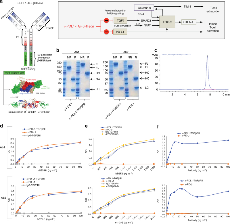
Fig. 6.
Design and bifunctional target binding ability of anti-PDL1-TGFβRII. a Schematic representation of PD-L1 and TGFβ entrained independent and cooperative mechanisms of immune tolerance. Schematic structure and targets of a-PDL1-TGFβRII are shown. a-PDL1-TGFβRII was designed by fusing the C terminus of the heavy chain of a human a-PD-L1 antibody with a ligand-binding sequence of the extracellular domain of TGFβ Receptor II (TGFβRII ECD) via a flexible linker peptide, (GGGGS)3. b SDS-PAGE under non-reducing (NR) and reducing (R) conditions was used to compare the full-length, heavy chain and light chain of a-PDL1-TGFβRII and a-PD-L1 antibody. Figure shows the results of SDS-PAGE analyses of each of two separate a-PDL1-TGFβRII Y-traps (Ab1 and Ab2) and their respective human a-PD-L1 antibody (atezolizumab and avelumab). c SEC-HPLC analysis of purified a-PDL1-TGFβRII; d ELISA showing the comparative ability of a-PDL1-TGFβRII and a-PD-L1 antibody to bind PD-L1. Biotinylated recombinant human PD-L1 (rh B7-H1-biotin; 0–100 ng ml−1) was added to plates coated with a-PDL1-TGFβRII or a-PD-L1 antibody (1 μg ml−1), followed by detection with HRP-Avidin. Plates coated with non-specific IgG-TGFβRII showed no binding to PD-L1 and served as a negative control to analyze the binding ability of the test samples. e ELISA showing the comparative ability of a-PDL1-TGFβRII and a-PD-L1 antibody to bind TGFβ. Recombinant human TGFβ (rhTGFβ1; 0–2,000 pg ml−1) was added to plates coated with a-PDL1-TGFβRII or a-PD-L1 antibody (1 μg ml−1), which was detected by biotinylated a-TGFβ1 and HRP-Avidin. Plates coated with nonspecific IgG-TGFβRII and rhTGFβRII-Fc served as positive controls to analyze the binding ability of the test samples to TGFβ. f ELISA showing the ability of a-PDL1-TGFβRII to simultaneously bind both PD-L1 and TGFβ. Anti-PDL1-TGFβRII or a-PD-L1 antibody (0–100 ng ml−1) was added to PD-L1-Fc coated plates (1 μg ml−1), followed by rhTGFβ1 (100 ng ml−1) that was detected by a biotinylated anti-human TGFβ1 antibody. For d–f, the data show the optical density (OD) values (mean of three replicate wells for each assay condition) from a representative of two independent experiments

