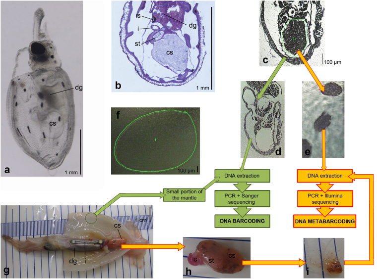
Figure 4.
Diagram of the lab workflow. (a–f) LCM gut content extraction (late paralarva E0 and early paralarvae, Table 3). (g–i) Direct dissection of gut contents (subadult and adult individuals E1 to E3 and late paralarvae E5 to E7, Table 3). (a) Lateral view of a live hatchling of the ommastrephid squid Todaropsis eblanae, obtained by in vitro fertilization (after Fernández-Álvarez et al.,15). (b) Histological sagittal section of a T. eblanae paralarvae, showing the structure of the digestive system. (c) Sagittal section of the early paralarva E41 (Dosidicus gigas) mounted on the PEN slide during a LCM session; the green line encircles the area selected for laser cutting. (d) Same section as in subfigure C with the caecum sac contents LCM-excised. (e) Cuts of LCM-isolated gut contents of several sections of the paralarva E41. (f) PEN slide without tissues (blank), the green line shows the portion selected for laser cutting. (g) Subadult individual E2 (Sthenoteuthis pteropus) with the mantle opened to show the internal organs. (h) Caecum sac and caecum of individual E2. (i) Isolated gut contents by direct dissection. Abbreviations: c, caecum; cs, caecum sac; dg, digestive gland; i, intestine; is, ink sac; st, stomach.

