Figure 4.

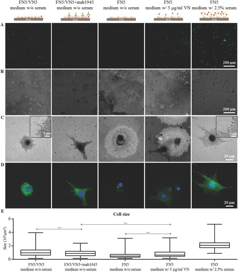
Cell‐mediated FN reorganization after 3 h incubation on FN/VN‐coated surfaces in serum‐free conditions. A) Actin cytoskeleton (green) and nuclei (blue). B) Corresponding cell‐mediated FN reorganization. C) Magnification of FN reorganization around single cells; insets indicate examples of FN reorganization by cells. D) Corresponding actin cytoskeleton (green) and nuclei (blue). E) Cell size. Mab1945 is used to block VN availability (second column); controls include reorganization of coatings of sole FN in serum‐free conditions (third column), with 5 µg mL−1 of VN in the culture medium (fourth column), and in the presence of 2.5% serum (fifth column). All differences are statistically significant (p < 0.05), with the exception of the ones indicated with ∼.
