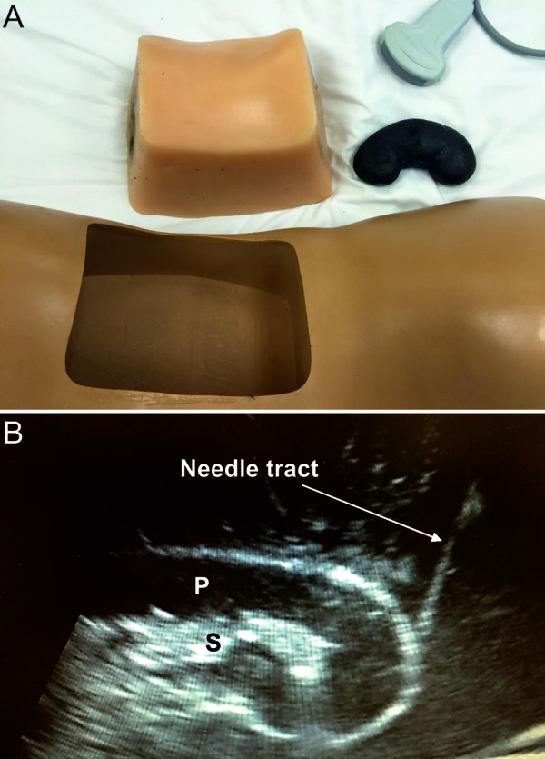
Figure 1. A: Display of the components of a commercially-available mannequin utilized in the fourth station of the Kidney Biopsy Simulation Workshop: insert of a model torso and a model kidney are shown. B: Image (sagittal view) obtained by ultrasound from a mannequin kidney (Blue Phantom™) during a real-time US-guided biopsy simulation, resembling the appearance of a native live kidney. Needle tract is depicted as it approaches the lower pole. S = renal sinus; P = renal parenchyma.

