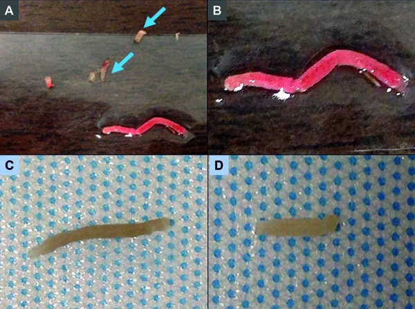
Figure 2. Images demonstrating appearance of kidney biopsy cores as shown to the Kidney Biopsy Simulation Workshop participants. A: Core biopsy of the renal cortex showing reddish-tan tissue. Fatty tissue in the periphery of the slide appears yellow (blue arrows). B: Higher magnification image of the kidney cortex. C: Kidney cortex after formalin fixation. D: Kidney medulla after formalin fixation.

