Figure 2.

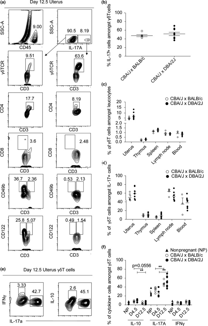
γδ T cells are the main producers of IL‐17A in pregnant mice. (a) Frequency of IL‐17A‐producing cells in the uterus of a representative pregnant BALB/c‐mated CBA/J female at day 12.5 of pregnancy. Numbers indicate frequency of the gated population. On the left, corresponding positive staining controls from CD45+ IL‐17A cells. On the right, IL‐17A+ cells. γδ TCR + CD3+ were used for γδ T cells. CD4 and CD8 were used in addition to CD3 cells to identify CD4 and CD8 T cells. CD49b and CD122 were used to identify NK cells, in addition to CD3 for NKT cells. (b) Percentage of IL‐17+ cells amongst γδ T cells in uteri of pregnant DBA/2J‐ or BALB/c‐mated CBA/J females on days 12.5 of pregnancy and of nonpregnant CBA/J females, n = 5–8 per group (c) Percentage of γδ T cells amongst CD45+ cells in various organs of pregnant DBA/2J‐ or BALB/c‐mated CBA/J females on day of 12.5 of pregnancy, n = 5–8 per group. (d) Percentage of IL‐17A‐producing γδ T cells (CD3+ γδTCR + cells amongst IL‐17+ cells) in various organs of pregnant DBA/2J‐ or BALB/c‐mated CBA/J females on day of 12.5 of pregnancy, n = 5–8 per group. (e) Frequency of cytokine‐producing‐γδ T cells (cytokine+ amongst CD3+ γδTCR + cells) in stimulated (with PMA and ionomycin) total uterine single cells of a representative pregnant BALB/c‐mated CBA/J female at day 12.5 of pregnancy. (f) Percentage of cytokine‐producing γδ T cells (IL‐10+, IL‐17+ and IFNγ+ cells amongst CD3+ γδTCR + cells) in uteri of pregnant DBA/2J‐ or BALB/c‐mated CBA/J females on days 4.5 and 12.5 of pregnancy and of nonpregnant CBA/J females, n = 3–10 per group. Results are shown as mean ± SEM and are representative of at least three experiments. Mann–Whitney test was used. *P < 0.05, **P < 0.01, ***P < 0.001. ▲ Nonpregnant, ○ CBA/J x BALB/c, ● CBA/J x DBA/2J.
