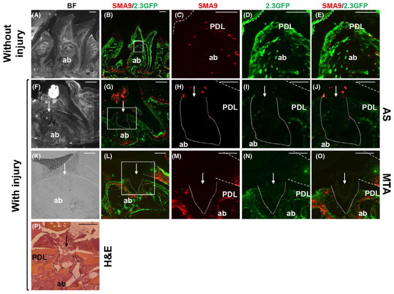
FIGURE 1.
Effects of experimental perforation of the integrity of periodontal ligament (PDL) and alveolar bone (ab). Representative images of sagittal sections through maxillary molars from αSMACreERT2;Ai9/Col2.3GFP mice. In all images the dental pulp is denoted by dashed lines, the site of injury by an arrow and the injury to the PDL and underlying ab by dotted lines. Bright-field (A) and epifluorescence (B) images of intact molars isolated 14 days after tamoxifen injection. (C–E) Higher magnification of boxed area shown in (B). Note the expression of SMA9+ cells (red) and 2.3GFP+ cells (green) in dental pulp, PDL and ab. Also note a few cells coexpressing SMA9/Col2.3-GFP (yellow) in PDL, indicating the differentiation of SMA9 into PDL fibroblasts. Bright-field (F, K) and epifluorescence (G, L) images of sections through injured maxillary molars filled with one-step self-etch Adhesive System (AS) (F–J) and mineral trioxide aggregate (MTA) (K–O) isolated 2 days after PDL injury. (H–J) Higher magnification of boxed area shown in (G). (M–O) Higher magnification of boxed area shown in (L). Note the lack of detectable SMA9+ and 2.3GFP+ cells at the sites of injury. Also note expansion of SMA9+ and 2.3GFP+ cells in bone marrow of the ab surrounding injury in molars restored with MTA (L–O). (P) Hematoxylin and eosin (H&E)-stained section of a maxillary molar isolated 2 days after injury (indicated by arrow). Note the destruction of dentin in the pulpal floor, PDL and ab at the site of injury. Also note residual dentin chips (*). Scale bars=100 μm

