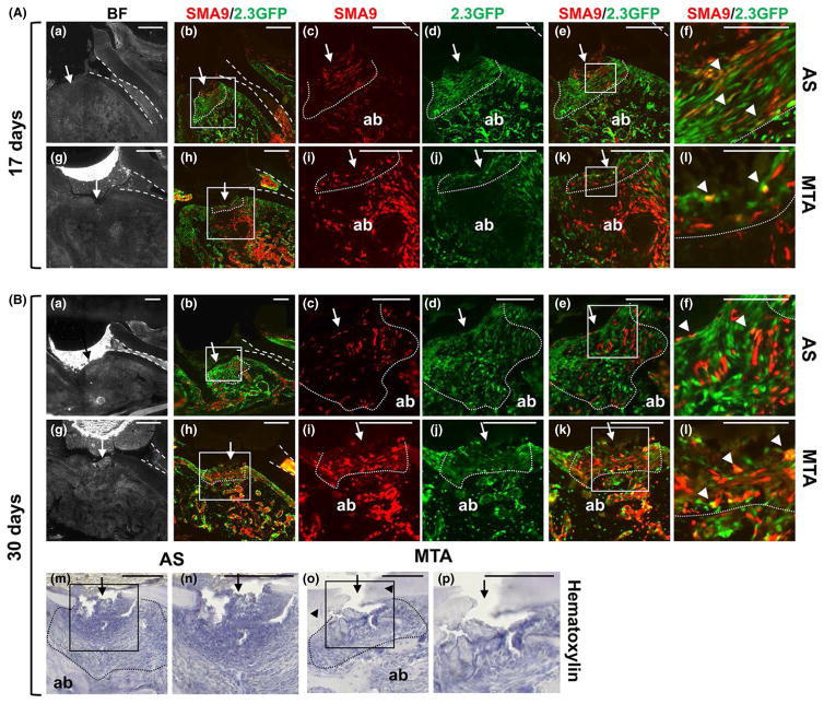
FIGURE 2.
Effect of mineral trioxide aggregate (MTA) on regeneration of periodontal ligament (PDL) and the underlying alveolar bone (ab). Representative bright-field and epifluorescence images of sagittal sections through injured maxillary molars from αSMACreERT2;Ai9/ Col2.3GFP mice. In all images the dental pulp is denoted by dashed lines, the site of injury by an arrow and the sites of repair by dotted lines. (A) Histology of injured molars 17 days after injury and restoration with one-step self-etch Adhesive System (AS) (a–f) and MTA (g–l). (c–f) Higher magnification of the boxed area in (b); (i–l) higher magnification of the boxed area in (h). SMA9+ cells are present in the PDL (outlined by dotted lines) and ab in both groups (c, i). A thick layer of cells expressing 2.3GFP are evident in AS-restored molars (d). Coexpression of SMA9 and 2.3GFP (yellow, arrowheads) is indicated. (B). Images of molars 30 days after injury and restoration with AS (a–f) and MTA (g–l). (c–f) Higher magnification of the boxed area in (b); (i–l) higher magnification of the boxed area in (h). Note the increase in number of SMA9+ cells in repaired PDL and ab in molars restored with MTA (i) than in molars restored with AS (c). Also note the increase in coexpression of SMA9+ and 2.3GFP+ cells (arrowheads) in repaired PDL of molars restored with MTA (f) than in molars restored with AS (l). Scale bar=50 μm (f and l) in A and 25 μm in B. (m–p) Hematoxylin-stained section of maxillary molars 30 days after injury (indicated by arrow). (n, p) Higher magnifications of boxed areas in (m) and (o), respectively. Repaired PDL and PDL region are outlined with dotted line. Note the lack of ab repair and large fibrous area containing fibroblastic cells in the PLD region in molars restored with AS (m, n). Also note well-organized repaired ab, PDL with PDL-like fibroblasts and partial dentin repair (arrowheads) in molars restored with MTA (o, p). Scale bar=100 μm

