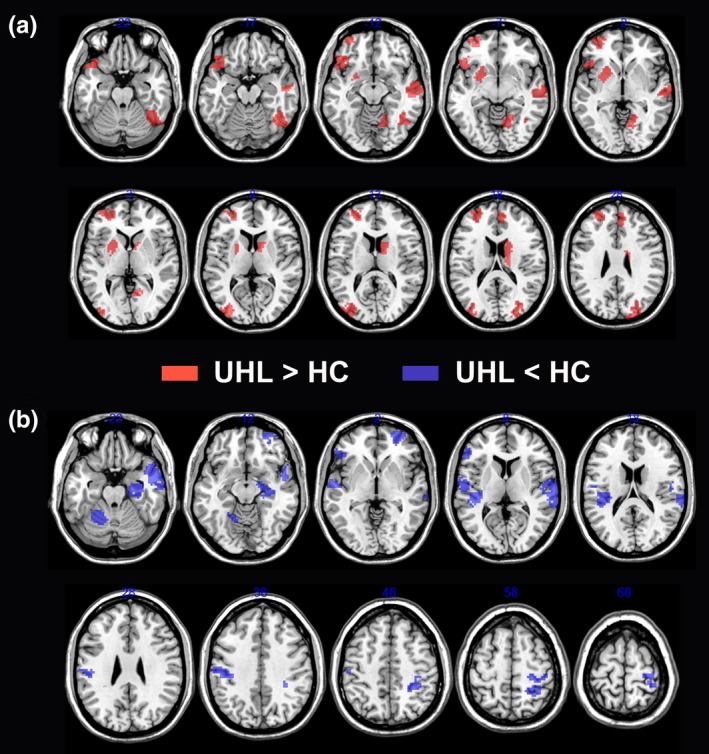
Figure 4.

Brain regions showing altered nodal betweenness centrality in brain functional networks. (a) UHL patients relative to the control subjects showed significantly increased nodal betweenness centrality. These regions were predominantly located in visual network, default‐mode network, and subcortical network. (b) UHL patients relative to the control subjects showed significantly decreased nodal betweenness centrality. These regions were mainly located in auditory network, visual network, default‐mode network, and attention network. HC, healthy control; UHL, unilateral hearing loss
