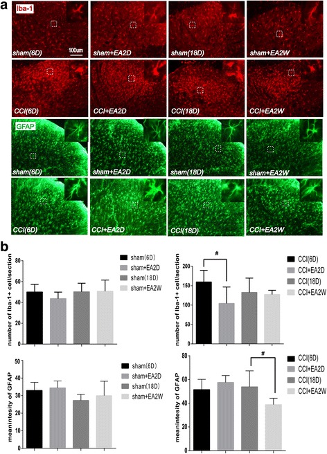
Fig. 4.

Immunofluorescence staining showing activities of microgliacytes and astrocytes in Ipsi and Cont DHs of lumbar spinal cord on day 6 and 18 after CCI in rats. a Representative tissue sections of immunofluorescence staining showing Iba-1-labeled microgliacytes (red) and GFAP-labeled astrocytes (green) in the Ipsi DHs of the lumbar spinal cord. In the superior-right corner of each section, the magnified microgliacyte and astrocyte from each dashed square box are shown. b Bar graphs showing the numbers of microgliacytes and mean fluorescence intensity values of GFAP (for astrocytes) in the Ipsi DHs on day 6 and 18 in the sham and EA groups(mean ± SD, n = 6/group). Results showed that in the early period of CCI-induced neuropathic pain, 2 days’ EA suppressed the activation of microgliacytes (not astrocytes), and in the later period, 2 weeks’ EA suppressed the activation of astrocytes. # P < 0.05, vs the CCI group)
