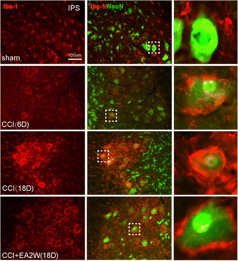
Fig. 5.

Representative section samples of dual immunofluorescent staining of Iba-1 (red) and neuronal nuclei (NeuN, green) for microgliacytes and neurons in the Ipsi ventral horns of lumbar spinal cord in the sham, CCI 6D and 18D, and CCI + EA2W groups. The images from the left to the right longitudinal rows are Iba-1-labeled microgliacytes, Iba-1 and NeuN labeled cells and magnified cells chosen from the dashed square box of each image on their individual left side. The NeuN-positive neurons were closely surrounded by many microgliacytes after CCI, suggesting a remodeling of the lumbar locomotor circuitry. NeuN, a neuronal specific nuclear protein and a biomarker for neurons
