Figure 3.

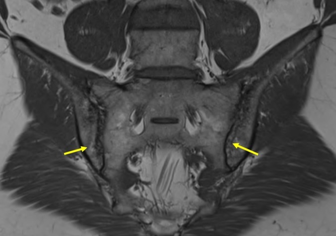
MRI, T1-weighted sequence of sacroiliac joints: presence of definite erosion in the ilium on both sides (arrows), perifocal fatty lesions of subchondral bone and localised sclerosis.

MRI, T1-weighted sequence of sacroiliac joints: presence of definite erosion in the ilium on both sides (arrows), perifocal fatty lesions of subchondral bone and localised sclerosis.