Figure 3.
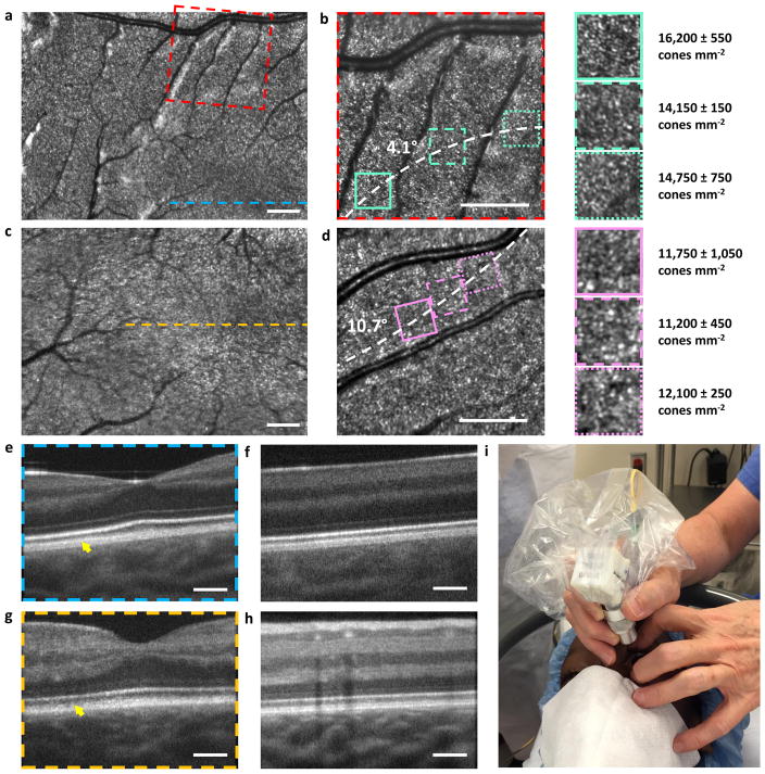
High resolution retinal images acquired on a healthy 25 month old toddler (a–b, e–f) and 14 month old infant (c–d, g–h). a, c, SLO images acquired near the fovea. b, d, Reduced FOV SLO images visualizing parafoveal photoreceptors. Zoomed insets (1.95×) and cone densities (mean ± one standard deviation as determined by three human graders) are shown to the right of b and d. e–h, OCT B-scans demonstrating similar image quality to that of the adult subject. i, photograph of the handheld probe in use on a child in the operating room. Dashed white curves in b and e pass through the centre of the zoomed insets at distances of 4.1° and 10.7° from the fovea, respectively. The yellow arrows in e and g indicate the interdigitation zone, which is well-defined in the 25 month old toddler but not fully developed in the 14 month old infant. Scale bars, 1°.

