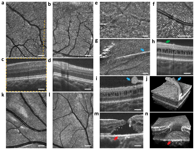
Figure 4.
Imaging results from children with eye disease: a 2 year old toddler with FEVR (a–d), a 2 year toddler with X-linked retinoschisis (e–j) and a 12 year old child with a macular hole (k–n). a–b, SLO images indicating undamaged regions of the macula. c–d, OCT B-scans with no visible abnormality as expected for this disease away from the peripheral retina. e–g, SLO images revealing fine membrane wrinkling of the retinal surface and an elevated, thicker surface membrane. h–j, OCT B-scans and volume showing retinal gaps characteristic of retinoschisis. Green arrow in h shows subtle wrinkling of the epiretinal membrane. Blue arrows in i–j reveal the elevated retinal membrane also visible in the SLO image in g. k–l, SLO images showing minimal damage to areas peripheral to the macular hole. m–n, OCT B-scan and volume showing the macular hole in microscopic detail with red arrows indicating corresponding sections of the hole.

