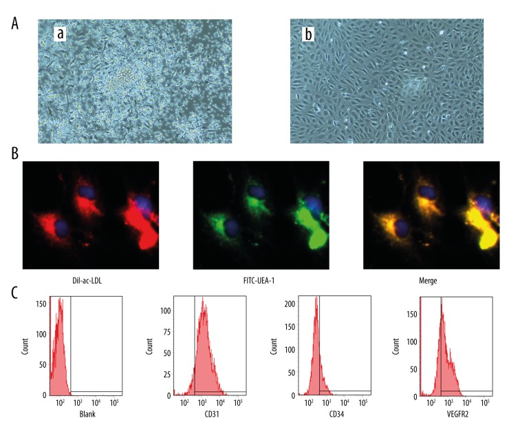
Figure 1.
Characterization of endothelial progenitor cells (EPCs). (A) EPCs colony exhibited a central cluster on day 4 (a) and formed a spindle shaped, endothelial cell-like morphology on day 14 (b) (100×). (B) Double staining of FITC-UEA-1 and DiI-ac-LDL and merge image (400×). (C) Expression of CD31, CD34 and CD309 of EPCs at day 14.

