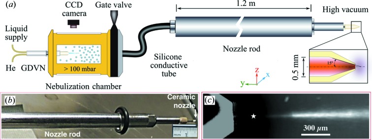
Figure 1.
CNAI assembly and its operation during the CXI experiment. (a) Sketch of the basic aerosol generation and transportation setup. (b) The aerosol nozzle mounted on the nozzle rod. (c) Time-integrated image of a laser-illuminated stream of GV particles exiting the CNAI, recorded using the in-line microscope at the CXI instrument. This image was formed by averaging over 3.7 min, with a running median background subtracted from each frame. The CNAI tip is seen in the left portion of the image, and the approximate X-ray focal point is indicated by the star.

