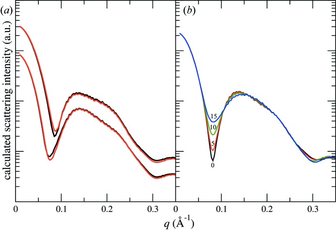
Figure 6.
Calculated scattering intensity from membrane-protein-loaded nanodiscs. The membrane protein is represented by a cylinder 44.3 Å in height. (a) Ambiguity in the bilayer structure discussed in §3.5.1 can lead to different scattering intensities expected for the same membrane protein located at the centre of the nanodisc. The calculations were done for two membrane protein diameters, 25 Å (bottom) and 45 Å (top). The empty nanodisc structures are taken from Fig. 5 ▸ with the same colour code. The intensities for the two different protein sizes are offset for clarity. (b) Varying the position of the membrane protein (35 Å in diameter) within the nanodisc results in dramatic changes in the scattering intensity. The same fluid-phase nanodisc structure (black lines) in panel (a) is used in this calculation. The membrane protein is systematically shifted along the major axis of the elliptical nanodisc from the centre by 0 (black), 5 Å (red), 10 Å (green) and 15 Å (blue).

