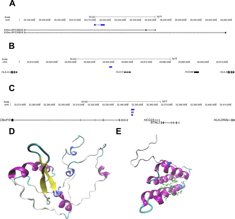
Figure 6.
Genomic position (hg19) and predicted protein structure of the three novel protein-coding genes identified in this study. (A) One protein-coding novel transcript (blue) contained within the intronic region of the gene XXbac-BPG308J9.3. (B) and (C) depicts novel protein coding transcripts (blue) in intergenic regions, near HLA-A and HLA-DRA, respectively. Each of these transcripts shares homology with endogenous retroviral elements. (D) The Predicted protein structure of transcript A (prediction p value= 1.17 × 10⁻⁴). This structure shares homology with an endogenous retroviral pol protein, and no predicted ligands are available. (E) The predicted protein structure of transcript C (prediction p value= 0.037). This structure shares homology with a retroviral gag protein, and is predicted to bind to a leucine residue (predicted active site amino acids are shown in green). In both D and E coloring is based on secondary structure: alpha helices are purple, 3–10 helices are blue, beta sheets are yellow, beta bridges are tan, turns are cyan, and coils are white.

