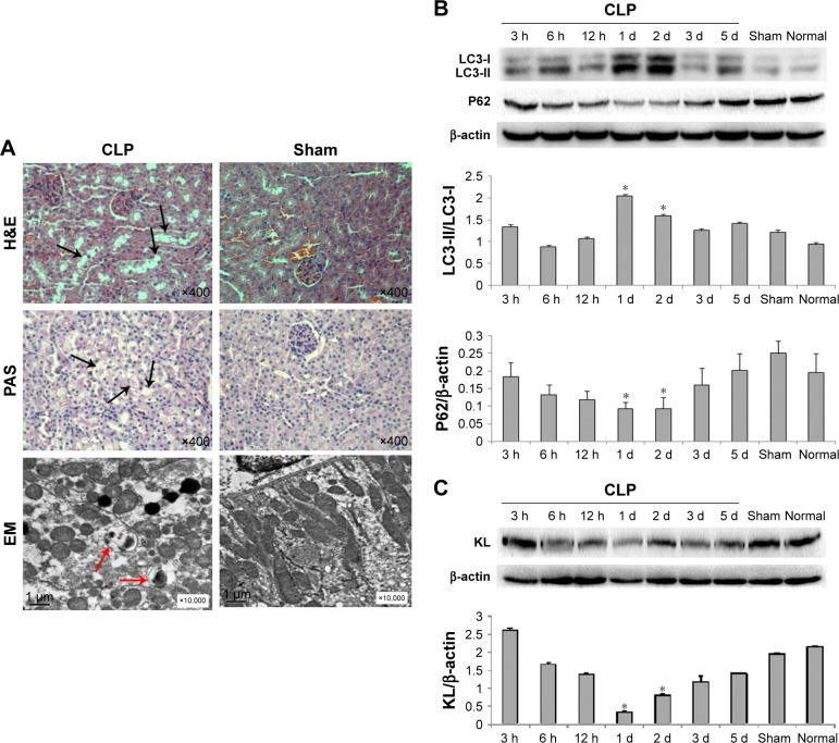
Figure 1.
Adverse effects of CLP on mouse kidneys.
Notes: (A) Images of H&E staining, PAS staining, and electron microscopy of kidney tissue 24 hours after CLP. Mouse kidney tissues were collected 24 hours after CLP and sectioned. Tissue sections were fixed and stained with H&E and PAS, which showed vacuolar degeneration and loss of brush borders (black arrows) in proximal tubular epithelial cells. Images at 400× magnification were acquired using a biological imaging microscope (BX53; Olympus Corporation, Tokyo, Japan). Tissue sections were also observed under a Hitachi H7500 electron microscope (Hitachi Ltd., Tokyo, Japan), which revealed engulfment of degraded cytoplasmic components by autophagosomes (red arrows). (B) Western blot for LC3-II, LC3-I, and P62. *P<0.05 vs the sham control. (C) Western blot for KL. *P<0.05 vs the sham control.
Abbreviations: CLP, cecal ligation and puncture; EM, electron microscope; PAS, periodic acid-Schiff; d, days; h, hours.

