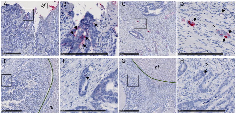
Fig. 2. F. nucleatum RNA ISH analysis of matched primary colorectal tumors and liver metastases.
Representative images of F. nucleatum spatial distribution in paired samples from P187 primary colorectal tumor (A and B) and liver metastasis (E and F) and P188 primary colorectal tumor (C and D) and liver metastasis (G and H) from the FFPE paired cohort are shown. Arrows indicate cells with histomorphology consistent with that of colon cancer cells infected by invasive F. nucleatum (red dots) in both primary colorectal tumors (B and D) and matched liver metastases (F and H). Fusobacterium-containing biofilm (bf) is highlighted in the colorectal tumor of P187 (A). Fusobacterium was not detected in normal liver (nl) tissue [(E) and (F)]. s, stroma. Panels (B), (D), (F), and (H) show magnification of the boxed areas in (A), (C), (E), and (G), respectively. Scale bars: 500 mm in (A), (C), (E), and (G); 250 mm in (B), (D), (F), and (H).

