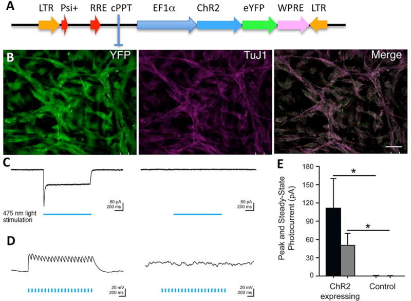
Figure 2.

NSCs express functional ChR2 transgene. (A) Schematic representation of the lentiviral vector carrying ChR2 fused to a fluorescent protein YFP and driven by the EF1a promoter (31). (B) Photomicrograph showing transduced NSCs differentiated into TuJ1+ neurons expressing the YFP reporter gene. (C) Sample voltage-clamp recordings show that 475 nm light (indicated by blue bar) induces inward photocurrents in NSCs stably expressing ChR2 (left) but does not produce a light-evoked response in control NSCs (right). Light power density is at 5 mW/mm2; cells were held at −65 mV. (D) Sample current clamp recordings show that blue light depolarizes NSCs expressing ChR2 (left), but not control cells (right). Light power density is at 5 mW/mm2; cells were held at −65 mV. (E) Summary bar graph of peak and steady-state photocurrent size. Mean ± SEM is plotted (n = 7) for ChR2-expressing cells (n = 5) for control cells. Scale bars: 10 μm (A).
