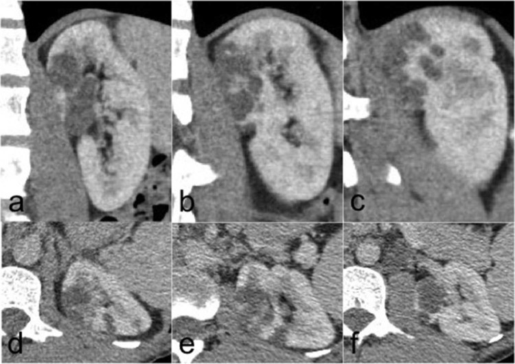
Fig. 2.
Sagittal (A-C) anterior to posterior and axial (D-F) upper pole to interpolar regions of the left kidney in the nephrographic phase of a triple-phase renal protocol computed tomography. Pre- and corticomedullary phases are not shown. This shows a focal intrarenal multiloculated cystic lesion in the anteromedial upper pole cortex.

