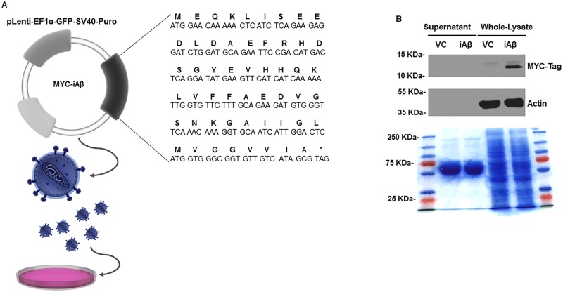
Fig 1. Validation of expression and localization of iAβ peptide.
(A) Schematic of the primary nucleotide and corresponding amino acid sequence of the MYC-Tagged iAβ used for cloning into the pLenti-EF1α-GFP-SV40-Puro lentiviral shuttle vector (Images adapted from Library of Science and Medical Illustrations CC BY-NC-SA 4.0). (B) Immunoblot of the concentrated supernatant and the whole cell-lysates of VC or iAβ transduced HEK293 cells for MYC-Tagged iAβ expression. Expression of the MYC epitope tag is observed in the whole cell-lysates of samples corresponding iAβ transduced cells only. Further, MYC-tagged iAβ is present in samples obtained from the whole cell-lysates and absent in samples obtained from the cultured supernatant confirming its intracellular retention. Coomassie brilliant blue stained, 12% SDS PAGE gel, of both concentrated supernatant and total cell lysates in VC and iAβ transduced cells demonstrates equal loading among samples.

