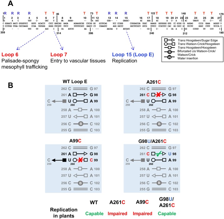
Fig 2. PSTVd RNA structures.
(A) The 2D organization of PSTVd RNA genome. 3D structural arrangements and the function of loop 6, loop 7, and loop E are listed [17–19]. “T” and “R” depict the functions in “trafficking” and “replication,” respectively [16]. (B) Disruptive and compensatory PSTVd loop E mutants predicted by isostericity [17]. Illustration for the replication of PSTVd variants in tomato plants, verified by northern blots [17], is shown in the lower panel. PSTVd, potato spindle tuber viroid; WT, wild-type.

