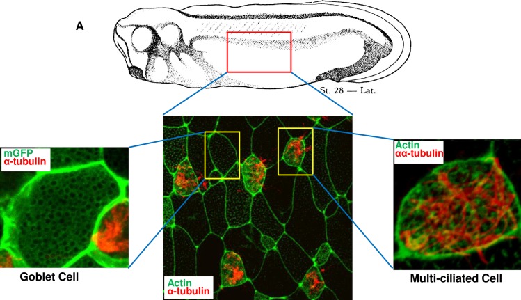
Fig 1. The composition of the muco-ciliary epithelium of Xenopus laevis embryo.
A. Immunofluorescence image of mucus secreting goblet cells and multiciliated cells in Xenopus embryonic epidermis. The membrane was stained using membrane GFP (green), Cilia were detected using α-tubulin (red) and actin by anti-actin antibody (green).

