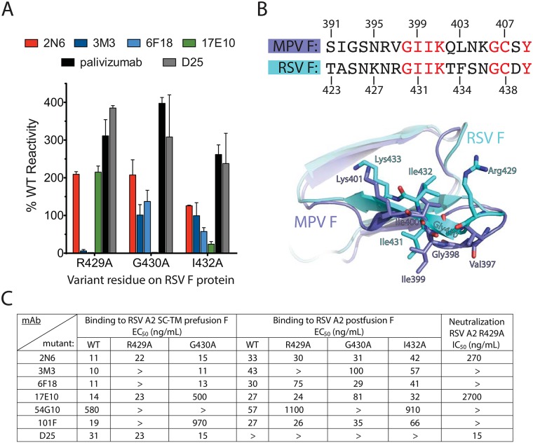
Fig 2. Characterization of antigenic site IV mutations.
(A) Alanine-scanning mutagenesis binding values for the generated site IV mAbs, compared with palivizumab and mAb D25 controls. The mAb reactivity for each RSV F variant was calculated relative to that of wild-type RSV F. Error bars indicate the measurement range of two independent experiments. (B) Overlay of RSV F and hMPV F sequences and crystal structures (PDB IDs: 3RRR, 5L1X, overlaid at chain H for each structure) at antigenic site IV, with RSV F residues from the alanine-scanning mutagenesis shown. Conserved residues between RSV F and hMPV F are displayed in red font. In the crystal structure overlay, RSV F residues are shown in cyan and hMPV F residues are shown in blue. (C) ELISA EC50 values for recombinant post-fusion or pre-fusion (SC-TM) mutant proteins for the site IV mAbs or controls. Neutralization IC50 values also are displayed for the RSV strain A2 F variant R429A.

