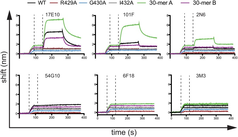
Fig 3. Binding curves determined by biolayer interferometry for mAbs targeting antigenic site IV.
Streptavidin biosensors were incubated with biotinylated peptides, and then mAbs were tested for binding in real-time. The first dashed line indicates the end of the peptide loading step, and the second dashed line indicates the beginning of the mAb binding step. After binding, mAbs were allowed to dissociate in real-time. The data are representative chromatograms from one experiment. Wild-type (WT) and mutant peptides consist of RSV F residues 422–436. The 30-mer A peptide includes RSV F residues 407–436, and the 30-mer B peptide includes RSV F residues 422–451.

