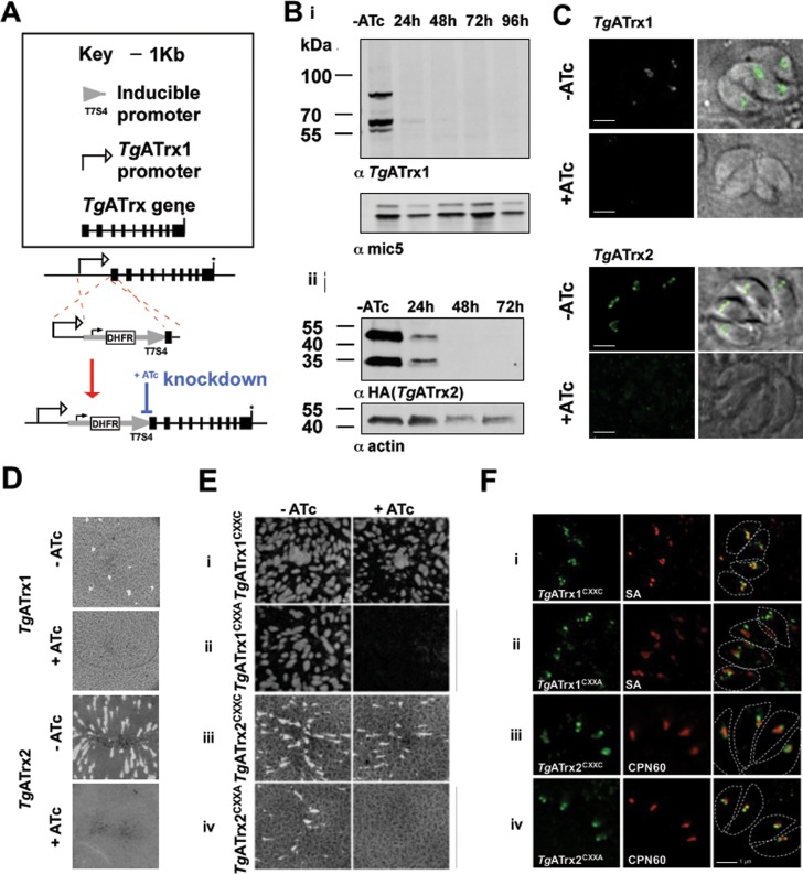
Fig 4. TgATrx1 and TgATrx2 are essential and their function requires their CXXC motifs.
A. Scheme of the manipulation performed to each of the TgATrxs loci to replace their promoters with the tetracycline-regulatable promoter. The gene model in the scheme is based on TgATrx1’s gene model, however the promoter replacement occurred via the same strategy for both TgATrxs. Black boxes–exons; asterisk–stop codon. B. Western blot analysis of TgATrx1 expression using anti-TgATrx1 (i), and endogenously HA-tagged TgATrx2 using anti-HA (ii), upon ATc treatment. C. Fluorescent microscopy showing TgATrx1 (anti-TgATrx1, bottom, green) and TgATrx2 (anti-HA, top, green) depletion at 48 hours of ATc treatment (+ATc) compared to non-treated control (-ATc). D. Plaque assays performed with TATiΔKu80PIATrx1 (top) and TATiΔKu80PIATrx2-3HA (bottom) with (+) or without (-) ATc. E. Plaque assays performed with TATiΔKu80PIATrx1 constitutively expressing a copy of TgATrx1CXXC (i) or TgATrx1CXXA (ii) and with TATiΔKu80PIATrx2-3HA constitutively expressing a copy of TgATrx2CXXC (iii) or TgATrx2CXXA (iv). F. Fluorescent microscopy of the localization of TgATrx1CXXC (i); TgATrx1CXXA (ii); TgATrx2CXXC (iii) and TgATrx2CXXA (iv), all in green, co-stained with Streptavidin (SA) which labels the apicoplast acetyl CoA carboxylase (i, ii) or CPN60 (iii, iv) both in red. White broken line shows parasites’ shapes. Scale bar, 1 μm.

