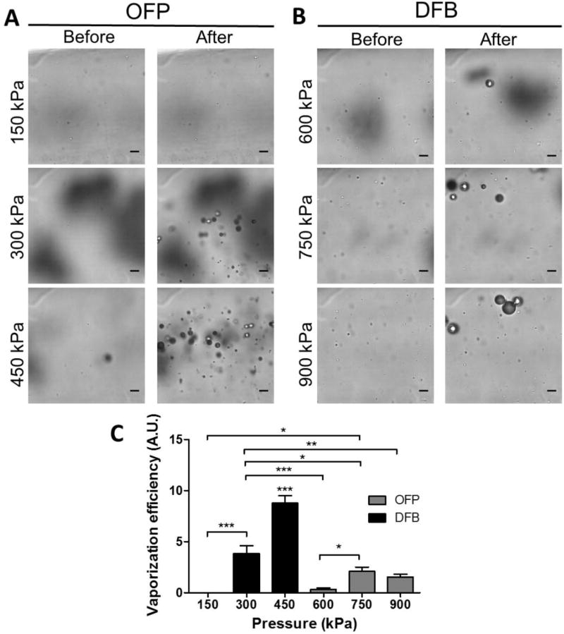
Figure 2.
Detection of acoustic droplet vaporization using high-speed optical microscopy (0 and 6 ms after sonication). (A) OFP-filled droplets were found to vaporize at pressures at and above 300 kPa, but not at 150 kPa. (B) DFB droplets were found to vaporize inconsistently at 600 kPa (vaporization did not occur with every activation pulse). Vaporization was consistently observed at 750 kPa and 900 kPa for DFB droplets. On average, more bubbles were generated from OFP droplets at low pressures (300–450 kPa) compared to those generated from DFB droplets at higher pressures (750–900 kPa). Scale bar represents 10 µm. (C) The relative vaporization efficiency was calculated by dividing the number of bubbles formed in the field of view by the nanodroplets concentration (1011 droplets/mL) measured with NanoSight. The vaporization efficiency of OFP droplets was higher than that of DFB droplets. Note that *** in (C) on top of OFP droplets at 450 kPa represents the same level of statistical significant to all the other cohorts.

