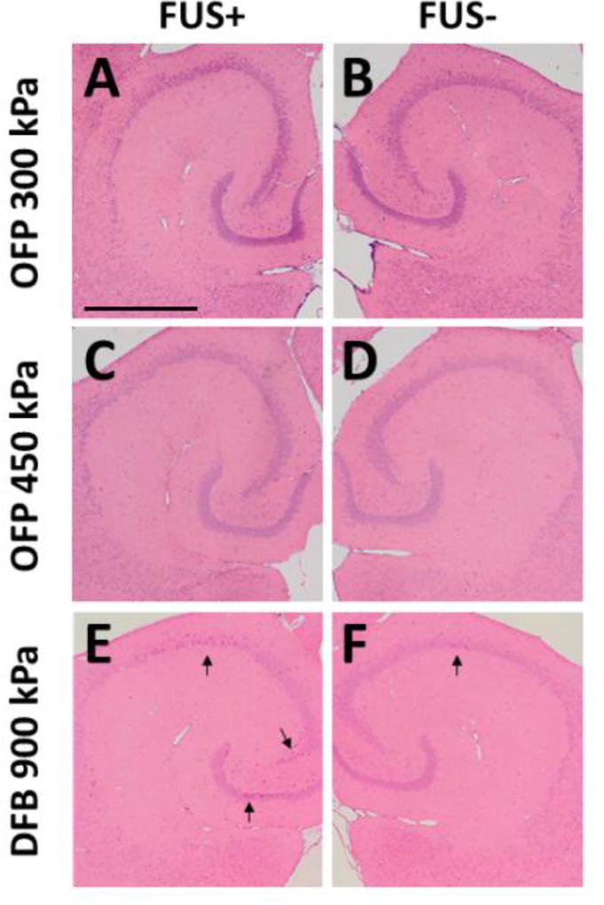
Figure 5.
Bioeffect assessment using histological staining (H&E). Sonicated (A, C, E) and nonsonicated (B, D, F) hippocampi using OFB at 300 kPa (A, B), 450 kPa (C, D), and DFB at 900 kPa (E, F). The results showed no damage (erythrocyte extravasations or dark neurons) using OFP droplets. For DFB droplets at 900 kPa, only 1 out of 3 animals showed an increased number of dark neurons on the hippocampi. The scale bar in (A) represents 1 mm.

