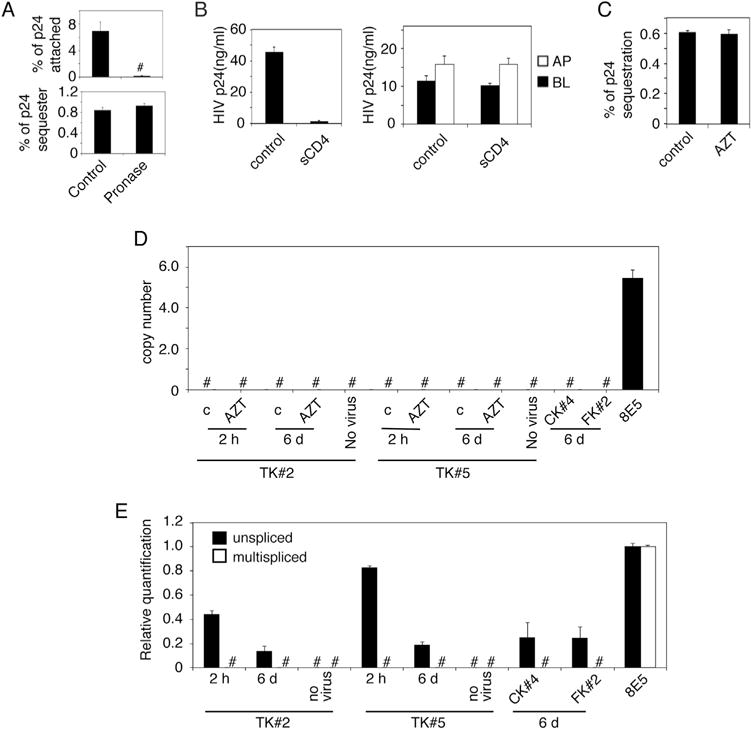
Fig. 7.

HIV-1 sequestration in polarized epithelial cells is not associated with membrane invagination and productive viral infection. (A) HIV-1SF33 was added to the AP surface of polarized tonsil epithelial cells for 1 h at 4 °C to determine viral attachment (upper panel). Another set of cells was used to determine sequestration of HIV-1SF33 for 6 days (lower panel). Cells with attached HIV-1 or sequestered HIV-1 were washed and treated with pronase. Untreated cells served as a control. Cells were collected and used for detection of attached and sequestered virions using ELISA p24. (B) HIV-1SF33 was added to the AP surface of tonsil cells and incubated at 4 °C for 1 h to allow virus attachment. sCD4 was added for 1 h at 4 °C (left panel). In parallel experiments, tonsil cells containing intracellular HIV for 6 days were exposed to sCD4 from AP and BL membranes for 1 h at 4 °C (right panel). Cells without sCD4 served as a control. Cells were washed and cocultivated with activated PBMC from AP membranes in cells with attached HIV and from AP or BL membranes in cells with sequestered virions for 4 h at 37 °C. PBMC were collected and cultured for 7 days and HIV infection was examined by ELISA p24. Data represent one of four independent experiments and are shown as mean ± SEM of triplicate values. (C) Polarized tonsil cells were pretreated with 10 μM AZT for 24 h, and HIV-1SF33 was added to the AP surface of cells for 4 h. Cells were washed, uninternalized virus was removed, and cells were cultured for 6 days with AZT. One set of cells with virus were untreated and served as a control. At day 6 cells were collected and examined for intracellular virus by ELISA p24. (D) Polarized tonsil TK#2 and TK#5 cells were treated with AZT for 24 h and exposed to HIV-1SF33. One set of cells were cultured for 2 h, and one set were cultured for 6 days. Polarized cervical CK#4 and foreskin FK#2 cells were exposed to HIV-1 and cultured for 6 days. As a positive control, we used an HIV-1-infected 8E5 T lymphocytic cell line. Cells were collected by trypsinization, and integration of HIV-1 DNA was examined using qPCR. (E) Polarized TK#2, TK#5, CK#4 and FK#2 cells were exposed to HIV-1SF33, and at 2 h or 6 days later, cells were collected and used for detection of unspliced and multispliced HIV-1 RNA by qPCR. Data represent one of three independent experiments and are shown as mean ± SEM of triplicate values.
