. 2018 Jan 26;7:e30391. doi: 10.7554/eLife.30391

Figure 5. Egr activation of the JNK pathway is required for the formation of ectopic wings.

(A) Frequency of ectopic wings (EW) following rnts>egr damage in wild type or CtBP334Δ4/+ (CtBP334Δ4 was generated via CRISPR/Cas9 on the same chromosome that contains rnts>egr) in listed genotypes: (1) hepr75/+, (2) co-expression of UAS-JNKDN and (3) co-expression of UAS-FosDN. (B) Frequency of ectopic wings (EW) following rnts> expression of listed transgenes (UAS-rpr, UAS-hepwt, UAS-hepCA, UAS-grindICD, UAS-ecto-egr) in wild type and CtBP-/+ genetic background. Temperature shifts for experiments shown in (A) and (B) were done on day 7 AEL for 40 hr. (C–H) Wing discs with homozygous mutant clones of FRT82B CtBPQ229* generated by mitotic recombination with the transcriptional reporters for AP-1-GFP (C, D), MMP1-lacZ (E, F) and dilp8-GFP (G, H).

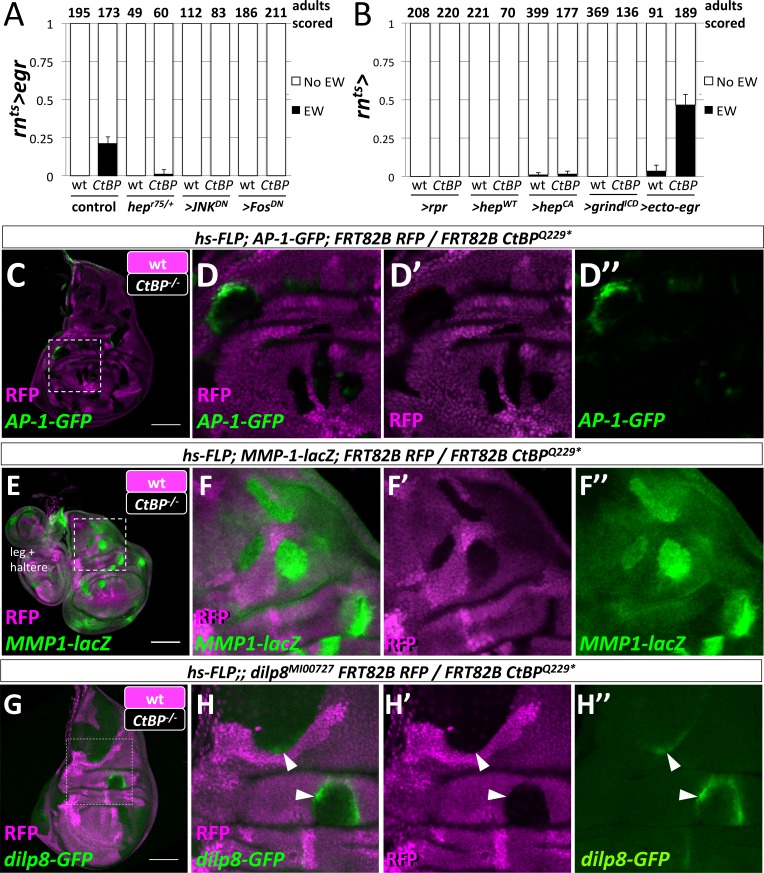
Figure 5.

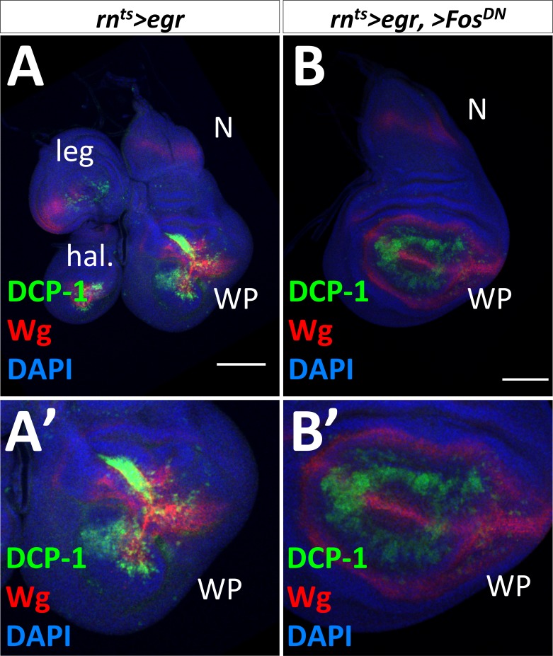
Figure 5—figure supplement 1. Co-expression of a dominant-negative Fos decreases the amount of tissue damage caused by rnts>egr.

Figure 5—figure supplement 1.

Wing imaginal discs following rnts>egr damage alone (A) or with UAS-FosDN (B). Note that in (B), the wing pouch (WP) has less apoptotic debris (DCP-1) and less Wg pattern disruption (compare B’) to A’). WP = wing pouch, N = notum, hal = haltere disc, leg = leg disc.
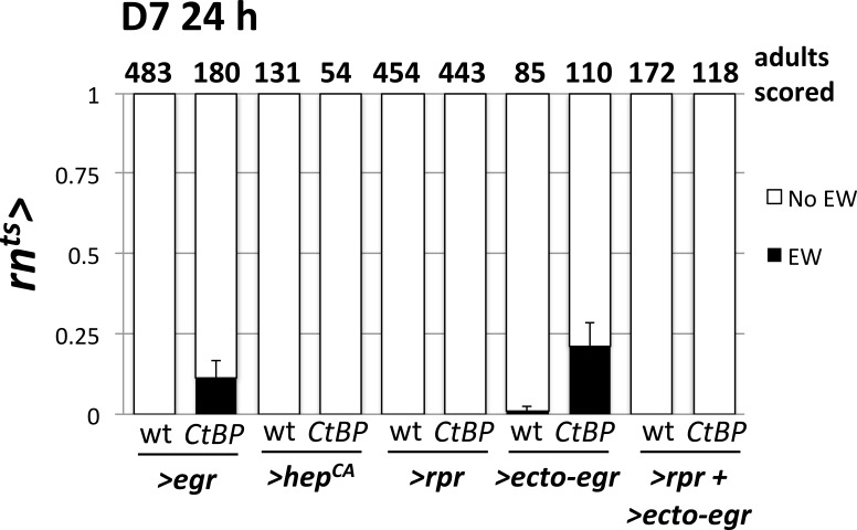
Figure 5—figure supplement 2. Shorter ablation period does not increase the frequency of ectopic wings.

Figure 5—figure supplement 2.

The frequency of ectopic wings (EW) following a 24 hr temperature shift on day 7 AEL in wild type and CtBPQ229*/+ genetic backgrounds. Damage was induced by rnts> driven expression of UAS-egr, UAS-hepCA, UAS-rpr, or UAS-ecto-egr alone, or by the co-expression of both UAS-rpr and UAS-ecto-egr. Compare EW frequencies to ones from 40 hr temperature shift, shown in Figure 2 and Figure 5. Both UAS-egr and UAS-ecto-egr show a decrease in EW frequency when shifted for a shorter period.
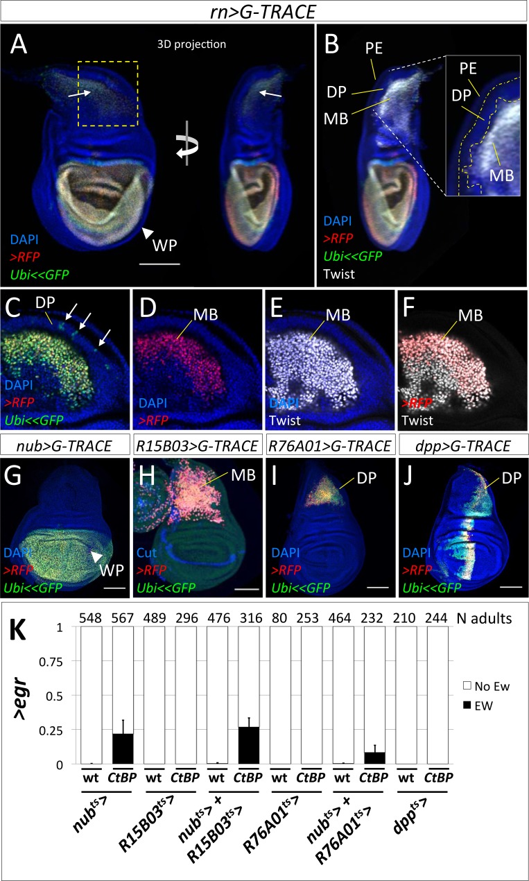
Figure 5—figure supplement 3. Expression of eiger in the wing pouch, but not in the myoblasts or notum epithelium, is sufficient to induce ectopic wings.

Figure 5—figure supplement 3.

(A, B) A 3D-projection of a late L3 wing disc of rn>G-TRACE, which shows current (UAS-RFPnls) and past (UAS-FLP, Ubi<stop<GFPnls) expression of rn-GAL4. There is strong rn> expression in the wing pouch (WP) and much weaker expression in a subset of the myoblasts (MB). (B) The same disc with MB marker Twist and a close-up to highlight the three cell layers in the notum: the peripodial epithelium (PE), the disc proper (DP) and the myoblasts (MB). (C–F) Close-up of the notum. (C) In addition to the MB, past expression (GFP) is detected in a few scattered cells in the DP of the notum (arrows). (D–F) Current expression (RFP) in the notum is limited to a subset of the MB, all of which express Twist. (G) nub>G-TRACE shows that current and past expression is mostly limited to the wing pouch (WP). (H) R15B03>G-TRACE shows expression is limited to the MB (anti-Cut identifies myoblasts and the stripe at the dorsoventral boundary of the DP). (I) R76A01>G-TRACE shows current and past expression is limited to the notum of the DP. (J) dpp>G-TRACE shows expression in the DP of the notum, the hinge, the WP and PE. (K) Frequency of EWs in adults following expression of >egr with the listed GAL4 drivers, in wild type or CtBPQ229*/+ genetic backgrounds. All cultures were shifted from 18°C to 30°C on day 7 AEL for 40 hr. Error bars show standard deviations.
Figure 5—figure supplement 4. Manipulations of apoptosis levels during the damage period.

Figure 5—figure supplement 4.

(A) Frequency of ectopic wings (EW) following rnts>egr damage in wild type or CtBP334Δ4/+ in listed genotypes: (1) Df(3L)XR38/+ and (2) co-expression of UAS-DroncDN. Same controls as shown in Figure 5A. (B) Frequency of EW following rnts>egr damage in wild type or CtBPQ229*/+ with the co-expression of UAS-p35, by transgenes inserted on chromosome 2 and 3. Same controls as shown in Figure 3J. (C, D) Wing imaginal discs following rnts>egr damage with co-expression of UAS-p35. Note that there is still a high level of apoptosis (DCP-1), and Wg pattern disruption (C) and JNK activity visualized by anti-MMP-1 (D). (E) Frequency of EW following rnts>egr damage with co-expression of UAS-rpr and UAS-ecto-egr. Shown same data as Figure 5B to compare to UAS-ecto-egr alone.
Figure 5—figure supplement 5. The regenerating wing pouch and ectopic pouch are derived from cells that express rn-GAL4 during the ablation period.

Figure 5—figure supplement 5.

(A) Diagram of the system used to lineage label cells that express rn-GAL4 during the ablation period. When shifted to the higher temperature (30°C), rn-GAL4 drives the expression of both UAS-egr and UAS-FLP. There are two independent FLP-out constructs (Ubi<stop<GFP and Act<stop<lacZ). There will be several possible classes of cells: (1) cells that do not express rnts>FLP and remain unlabeled, (2) cells that express low levels of rnts>FLP during the temperature shift and stochastically activate one construct (either express GFP or β-GAL alone), and (3) cells that express high levels of rnts>FLP and activate both flip-out constructs (express GFP and β-GAL). (B–I) Wing discs following 72 hr of recovery from rnts>egr ablation with an ectopic pouch (B–E) and without an ectopic pouch (F–I), as shown by pouch marker Pdm2. The wing pouch (WP) is largely regenerated from cells that expressed rnts>egr and UAS-FLP during the temperature shift. The ectopic pouch (EP) and notum (N), as highlighted in dotted rectangle in (D, H) are enlarged in (E, I) to show the classes of cells present. Green arrow = GFP only, Blue arrow = β-GAL only, White arrow = GFP and β-GAL and Red arrows are unlabeled regions that express Pdm2.
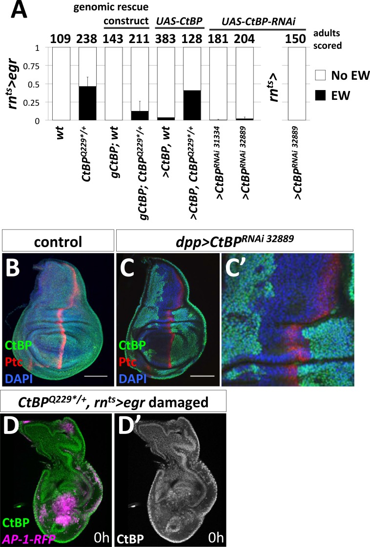
Figure 5—figure supplement 6. CtBP acts outside of the rn-GAL4 domain to prevent damage-induced ectopic wings.

Figure 5—figure supplement 6.

(A) Frequency of ectopic wings (EW) following rnts>egr damage as calculated by the fraction of adults without (no EW) and those with one or two ectopic wings (EW) from multiple biological replicates in listed genetic backgrounds. gCtBP is a genomic rescue construct that is located at attp40 (Zhang and Arnosti, 2011). Last column is rnts> control for >CtBPRNAi. Error bars show standard deviations. Numbers above bars represent the total number of adults scored for each genotype. (B–C) Wing imaginal discs stained with antibodies against CtBP and Patched (Ptc) in control (B) and with dpp-GAL4 driving the expression of UAS-CtBPRNAi (BL:32889) (C). CtBP immunoreactivity is lost in cells that currently express dpp-GAL4, which significantly overlaps with Ptc expression domain, as well as more anterior cells that expressed it earlier in development (likely due to RNAi persistence [Bosch et al., 2016]). (D) Wing disc following rnts>egr damage in CtBPQ229*/+ genetic background with AP-1-RFP reporter showing activation in the notum, stained with anti-CtBP. Note relatively uniform expression of CtBP (D’).
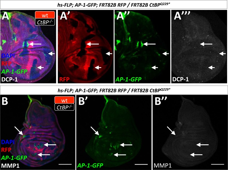
Figure 5—figure supplement 7. CtBP-/- mutant clones upregulate AP-1-GFP expression without evidence of apoptosis.

Figure 5—figure supplement 7.

(A–B) Wing disc with homozygous CtBP-/- mutant clones, which are RFP negative with the AP-1-GFP transcriptional reporter. (A) Disc stained for apoptosis (DCP-1). (B) Disc stained for MMP1. Note that there is clear AP-1-GFP reporter activation in CtBP-/- mutant clones, but without detectable apoptosis or MMP1 protein (arrows).