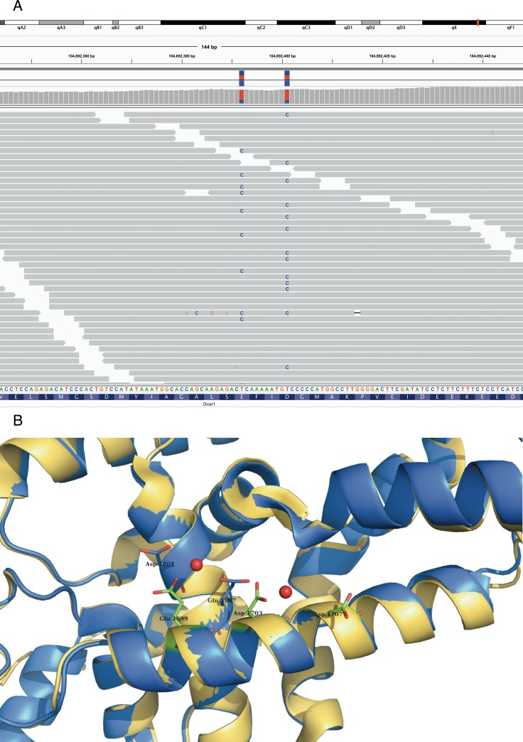
Figure 3. Bi allelic Dicer1 mutation.
Bi-allelic Dicer1 mutation A. IGV view of DICER1 mutated residues in tumour case 20: all but one read (with multiple mismatches and low mapping quality) show only one mismatch which indicates a bi allelic mutation in this tumor. B. mutated residues which are shown as sticks (in blue) are located at the surface of the protein, and bind together with Glu1699, Asp1703 and Asp1707 Mg (red balls). PDB-entry 3C4B [58] is shown in blue, PDB-entry 2EB1 [60] is shown in yellow.

