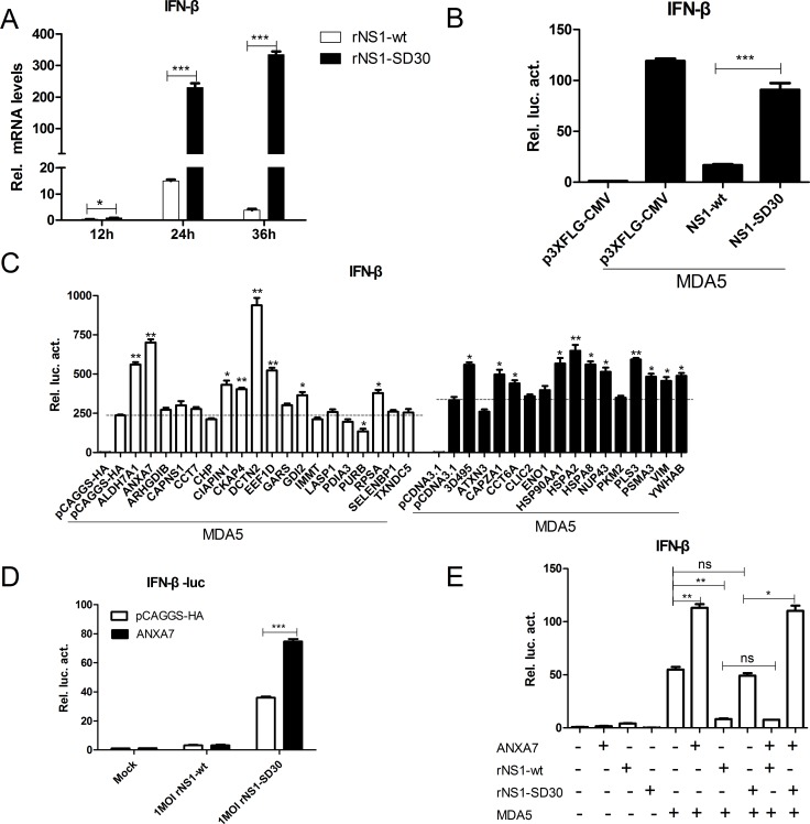
Figure 8. ANXA7 promotes the IFN-β response.
(A) mRNA expression levels in CEFs infected by rNS1-wt and rNS1-SD30 viruses. Uninfected and rNS1-wt- and rNS1-SD30- infected CEFs (0.01 MOI) were collected at 12, 24, and 36 hpi. Total RNA extracted from mock and virus-infected cells was used to detect chicken IFN-β mRNA by qRT-PCR. (B) NS1-wt inhibits IFN-β promoter activity induced by chMDA5. DF-1 cells were transfected with chMDA5 (100 ng) or empty vector (100 ng) and the indicated expression plasmids (200 ng) together with the pGL3-chIFN-β reporter plasmid (100 ng) and the pRL-TK vector (10 ng) for 24 h. Cells were lysed prior to luciferase assays; expression results reflect comparison to luciferase activity in control cells. (C) Effect of proteins overexpress on chMDA5 induced IFN-β response. DF1 cells were transfected with the indicated expression plasmids (200 ng) for 24 h, and subjected to luciferase assays as described in (B). (D) ANXA7 promotes IFN-β promoter activity induced by rNS1-SD30 virus. DF-1 cells were transfected with the indicated plasmids for 24 h, and subsequently infected with 1 MOI rNS1-wt or rNS1-SD30 virus or left untreated for 24 h. Luciferase assay was performed as described in (B).

