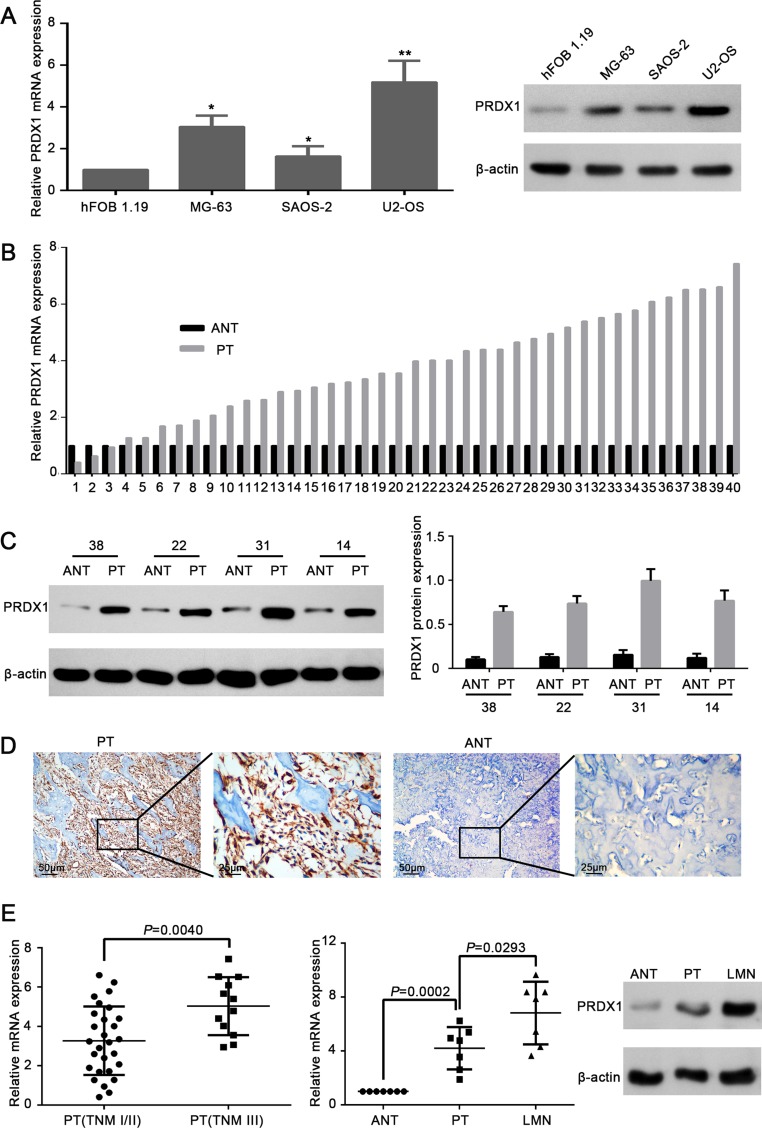
Figure 1. Expression level of PRDX1 elevates in human osteosarcoma tissues and cell lines.
(A) PRDX1 was overexpressed in osteosarcoma cell lines. Expression level of PRDX1 increased significantly in osteosarcoma cell lines MG-63, SAOS-2 and U2-OS when comparing to hFOB 1.19(normal osteoblast cells. Expression level was examined by qRT-PCR (left panel) and western blot (right panel). (B) qRT-PCR showed the mRNA level of PRDX1 was higher in PTs than in corresponding ANT. PT, primary tumor tissue; ANT, adjacent non-tumor tissue (n = 30). (C) Representative images of Western blot. Results showed the protein expression level of PRDX1 in PTs was higher than in corresponding NCBTs (n = 30). (D) Representative images of immunohistochemistry (IHC) staining of PRDX1 in osteosarcoma tissues. PTs exhibited strong positive signal than corresponding ANT. Magnification of images, × 400. (E) PRDX1 mRNA level was higher in TNM stage III (n = 12) than in TNM stage I/II (n = 30). (F) PRDX1 level was increased both at mRNA (left panel) and Protein level (right panel) in LMNs than corresponding NCBTs from the same patient (n = 7). LMNs, lung metastatic nodules.

