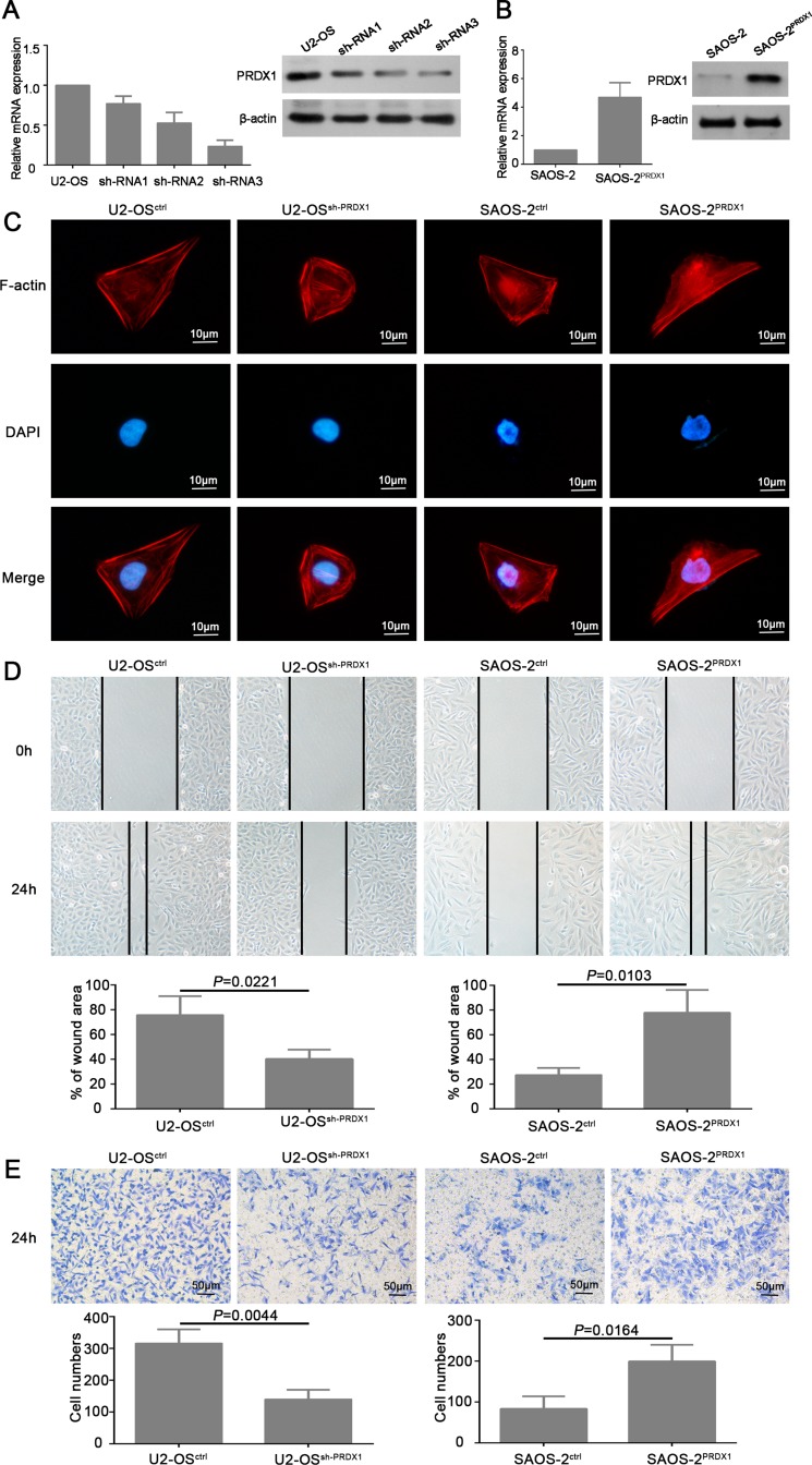
Figure 3. PRDX1 promotes migration and invasion of osteosarcoma cells in vitro.
(A and B) Establishment of PRDX1 knockdown and overexpression stable cells line. U2-OS was transfected with shRNA plasmids (A) and PRDX1 overexrpession plasmids (B). PRDX1 expression level was examined by qRT-PCR and western blot after successfully generating stable cell line. (C) Manipulation of PRDX1 changes cell morphology of osteosarcoma cells. F-actin filaments were visualized by using rhodamine-phalloidin. (D) Wound-healing assays showed that knockdown of PRDX1 suppress cell motility and invasion when comparing with control cells (left panel). Meanwhile, overexpression of PRDX1 promotes wound healing capacity when comparing with control cells. (E) Transwell assays showed that knockdown of PRDX1 suppresses cell migration when comparing with control cells (left panel). Meanwhile, overexpression of PRDX1 promotes cell migration in comparing to control cells.

