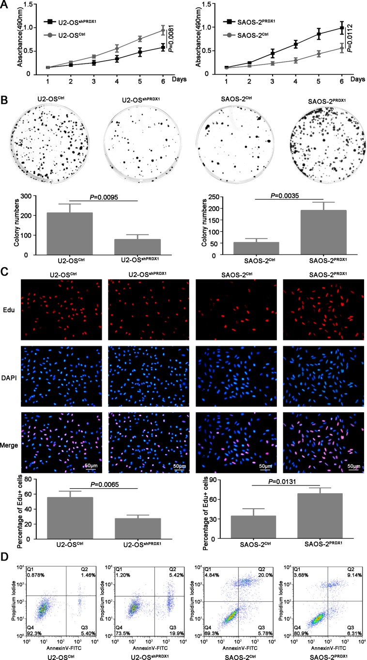
Figure 4. PRDX1 promotes proliferation of osteosarcoma cells in vitro.
(A) MTT was used to measure the proliferation ability of U2-OSshPRDX1 and U2-OSControl, SAOS-2PRDX1 and SAOS-2Vector cells. (B) The colony formation assay shown that knockdown of PRDX1 inhibits colony formation (left panel) and overexpression of PRDX1 promotes colony formation (right panel). (C) Cells were labeled with EdU for 3 hrs and the results revealed that the EdU positive cells were higher in osteosarcoma cells with relative higher PRDX1 level. (D) Cell apoptosis was measured by PI/Annexin V. Knockdown of PRDX1 induce cell apoptosis (left panel) but overexpression of PRDX1 suppress cell apoptosis (right panel).

