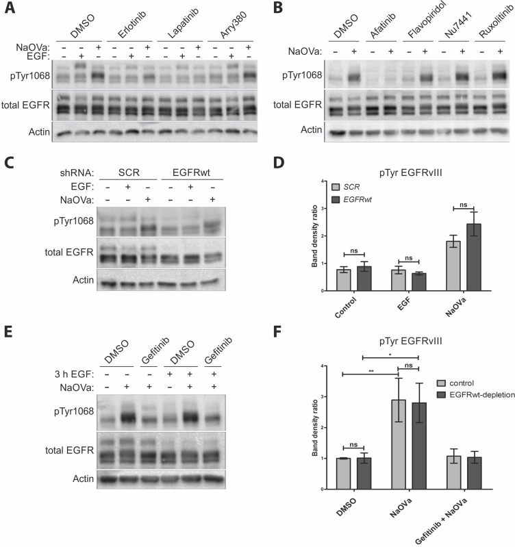
Figure 3. EGFRvIII is not phosphorylated by EGFRwt.
(A) Western blot analysis of cells treated with indicated inhibitors prior to and concurrently with EGF or NaOVa treatment for 1 h. Concentrations of inhibitors were based on literature reports. (B) Cells were treated with inhibitors specific to pan-JAK (ruxolitinib), CDK2 (flavopiridol) or DNA-PK (Nu-7441) prior to and concurrently with NaOVa treatment. Concentrations of inhibitors were based on literature reports. (C) Cells transiently expressing control scrambled (SCR) shRNA or shRNA targeted against EGFRwt were treated as indicated and analyzed by Western blotting. (D) Quantification of blots as shown in C, with columns representing ratio of phosphorylated to total EGFRvIII protein. (E) Cells treated with cycloheximide and stimulated with EGF for 3 h as indicated. Where indicated, cells were incubated for another 30 min with DMSO or gefitinib prior to 1 h treatment with NaOVa. (F) Quantification of blots as in E for EGFRvIII, with normalization to DMSO treated Control cells. Statistical analysis performed using two-way ANOVA with post-analysis Bonferroni’s multiple comparisons test. **p < 0.01; *p < 0.05; ns – not significant; n of at least 3 experiments. Error bars indicate SEM. Immunoblots have been uniformly adjusted for brightness and contrast to facilitate interpretation.

