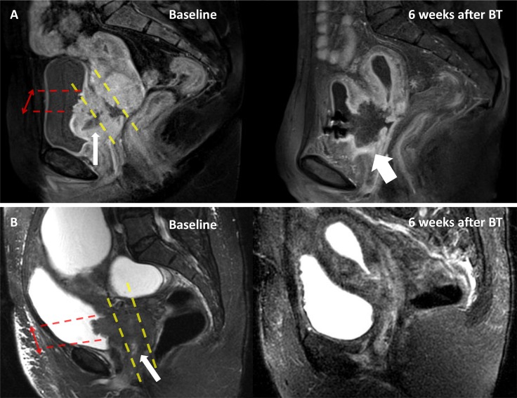
Figure 2. Example of anterior necrosis leading to VVF.
Magnetic Resonance Imaging (MRI) exams of two patients with cervical cancer stage IVA (patient A: contrast enhanced T1-weighted MRI and patient B: T2-weighted MRI) at baseline (left), and 6 weeks after brachytherapy (right). Patient A had a tumor necrosis (thin arrow) which involved the anterior third of the tumor, while there was no anterior necrosis for patient B. Six weeks after brachytherapy (BT), patient A developed a vesicovaginal fistula (thick arrow), while patient B had a complete response. Red arrows: height of the bladder involvement. Yellow dashed line: delimit the anterior-third, mid-third and posterior-third of the tumor.

