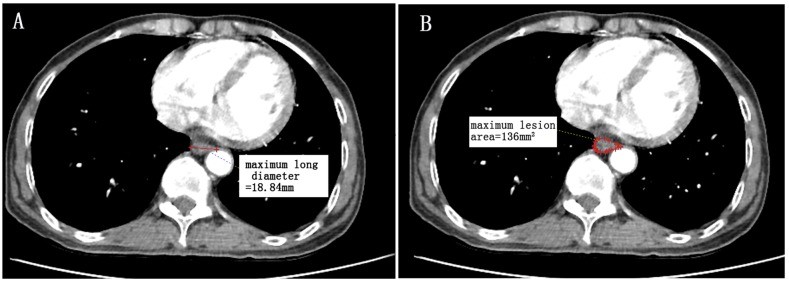
Figure 9. A patient with lower thoracic esophagus squamous cell carcinoma (T1N0M0).
An illustration of radiographic factors and endoscopic pathology reports that the distance from the incisors to the proximal edge of the tumor is 36cm. (A) maximum long diameter (18.84mm), (B) maximum lesion area (136mm2). Since no wall thickness was greater than the 5mm level as determined by CT based diagnostic criteria, CT-based lesion length is considered to be 0mm. This patient is “iT1”. (Measured by Neusoft PACS/RIS version 3.1,Neusoft Beyond Technology).

