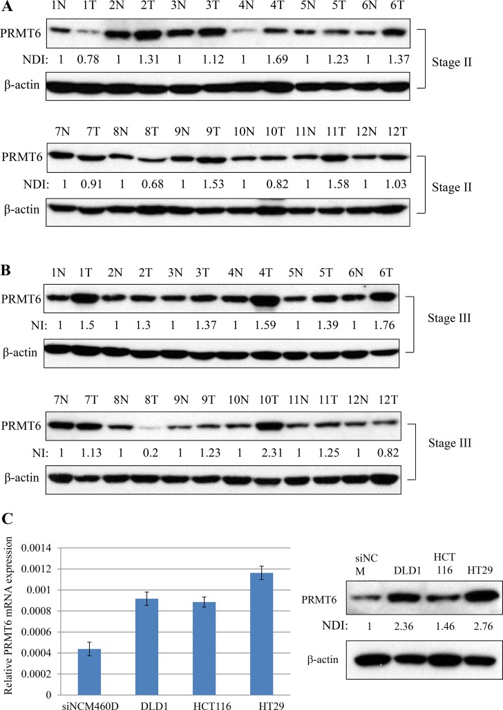
Figure 1. Overexpression of PRMT6 in CRC tissues and cell lines.
(A) Comparison of PRMT6 expression between the primary CRC and adjacent normal tissues paired from the same patient with stage II. (B) The same experiments in panel (A) were carried out with tissue extracts from the patients with stage III. (C) PRMT6 mRNA and protein expression levels between NCM460D and three CRC cells were compared by real-time PCR (left panel) and western blotting (right panel), respectively. Densitometric intensity of PRMT6 protein was normalized to β-actin. NDI indicates normalized densitometric intensity.

