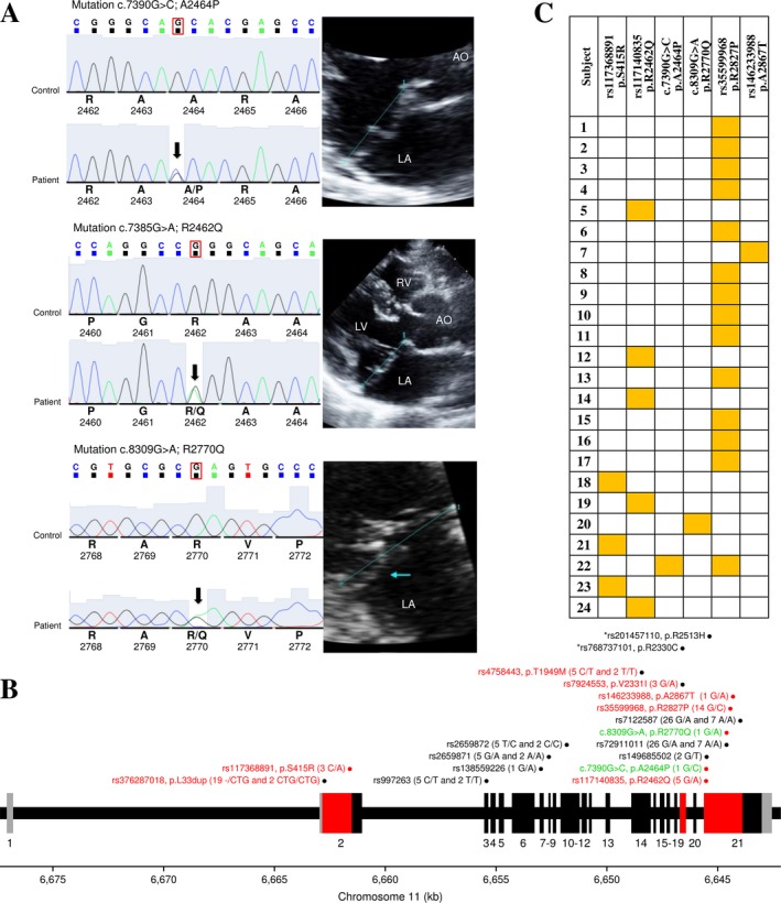
Figure 1.

Identification and characterization of genetic variants in the DCHS1 gene in patients affected by mitral valve prolapse. (a) Sequence chromatograms of the novel (p.A2464P) and rare (p.R2462Q and p.R2770Q) missense in silico‐predicted deleterious variants identified in this study with two‐dimensional echocardiographic long‐axis view of a representative heterozygote patient for each variant. The blue line denotes the mitral annulus. (b) The exon–intron structure of the DCHS1 gene and the localization of the identified genetic variants. The coding exons are shown in black (or red) and the untranslated regions in gray. The regions of the gene sequenced among the 100 patients are in red. Genetic variants are illustrated with their rs numbers if available with genotyping counts in parentheses for 12 or 100 patients. Red dots illustrate the six in silico‐predicted deleterious variants. The two functional variants identified by Durst et al. (2015) on exons 19 and 21 are illustrated on top in black with an asterisk. Missense and synonymous variants identified in this study are indicated in red and black, respectively. Newly and rare identified variants with no rs number are illustrated in green and named based on standard gene mutation nomenclature (den Dunnen et al., 2016). (c) Summary of patients carrying at least one of six variants identified and considered deleterious in this study. Heterozygote carriers are identified by a yellow box
