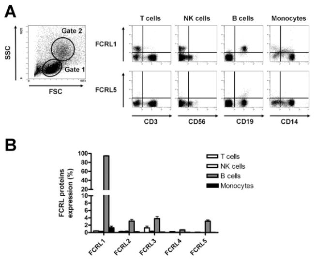
Figure 1.
Fc receptor–like (FCRL) protein expression by fresh peripheral blood mononuclear cells (PBMCs) from healthy donors. PBMCs from 20 healthy donors were stained with phycoerythrin-conjugated anti-FCRL monoclonal antibody and antibodies against lineage-specific markers. A, Left, Cells in the lymphocyte light-scatter gate (gate 1) were analyzed for CD3+ cells (T cells), CD56+ cells (natural killer [NK] cells), and CD19+ cells (B cells), while cells in the gate with larger scattering parameters (gate 2) were analyzed for CD14+ cells (monocytes). Right, Representative dot plots show expression of FCRL-1 and FCRL-5 on PBMCs from a healthy donor. FCRL-1 was expressed on the B cell fraction (CD19+). FCRL-5 was expressed on a small subpopulation (<4%) of B cells. B, The percentage of cells expressing FCRLs 1–5 was determined in the T cell, NK cell, B cell, and monocyte fractions. Bars show the mean ± SD.

