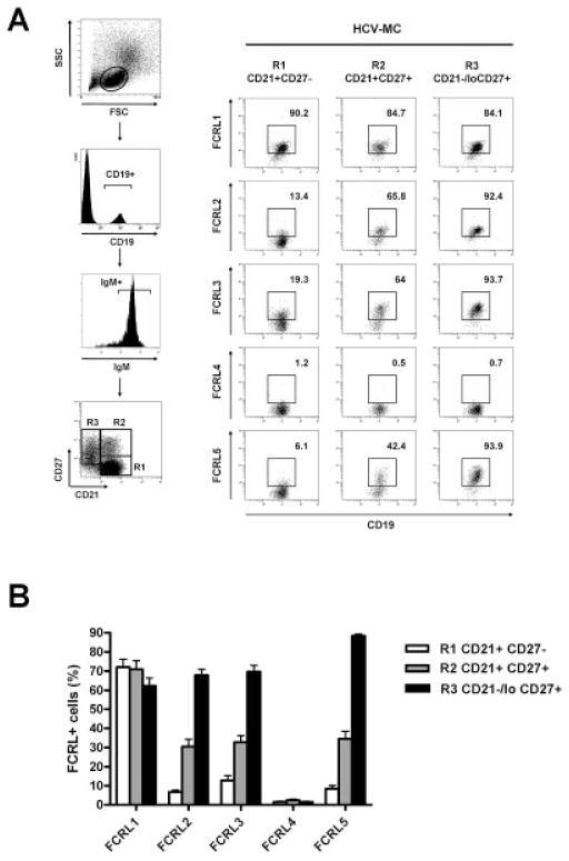
Figure 3.
Pattern of FCRL protein expression by naive B cells, conventional CD21+ B cells, and clonal CD21−/low MZ B cells from patients with HCV-MC vasculitis. PBMCs from 15 patients with HCV-MC vasculitis were stained with PE-conjugated anti-FCRL proteins, fluorescein isothiocyanate–conjugated anti-CD21, energy-coupled dye–conjugated anti-CD19, allophycocyanin–conjugated anti-IgM, and PE–Cy7–conjugated anti-CD27. After gating on CD19+ and IgM+ cells, CD21+CD27− naive B cells (R1), CD21+CD27+ MZ B cells (R2), and clonal CD21−/low MZ B cells (R3) were gated for analysis of expression of FCRL proteins 1–5. A, Representative dot plots show gating strategy (left) and expression of FCRL proteins 1–5 by naive B cells, conventional CD21+ MZ B cells, and clonal CD21−/low MZ B cells (right) in a patient with HCV-MC. B, Expression of FCRL proteins 1–5 by naive B cells, conventional CD21+ MZ B cells, and clonal CD21−/low MZ B cells was assessed in all patients with HCV-MC vasculitis. Bars show the mean ± SD. See Figure 2 for definitions.

