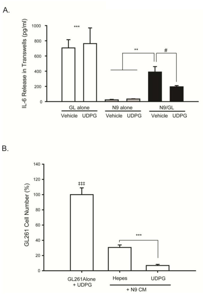
Figure 4. Pharmacologic activation of P2Y14 receptors with UDPG decreases IL-6 release in N9 microglia and GL261 glioma transwell cultures, and conditioned medium from microglia treated with UDPG decreases GL261 tumor cell proliferation.
(A) ELISA assays were performed to measure IL-6 concentrations in the tissue culture medium from GL261 tumor cells alone, N9 microglia alone, or from N9/GL261 transwell cultures grown together for 24 hours treated with either vehicle (250 mM HEPES) or UDPG (300 μM). GL261 cells produced the most IL-6, but the presence of microglia in transwell culture reduced this, an effect that was further reduced by treatment with UDPG. # p=0.09, ** p<0.015, and *** p<0.001 GL alone vs. all others. (B) N9 microglia were cultured alone and treated with either vehicle (250 mM HEPES) or UDPG (300 μM) for 24 hours. The N9 cell conditioned medium (N9 CM) was filtered and then applied to GL261 cell cultures at a ratio of 1:1 for 24 hrs after which time, cell numbers were quantified using the MTS assay. N9 CM data are expressed relative to GL261 cells treated directly with UDPG to account for the presence of UDPG in the microglia conditioned medium. CM from UDPG-treated N9 cells reduced GL261 glioma cell proliferation. *** p < 0.001 vs. GL261 cells alone.

