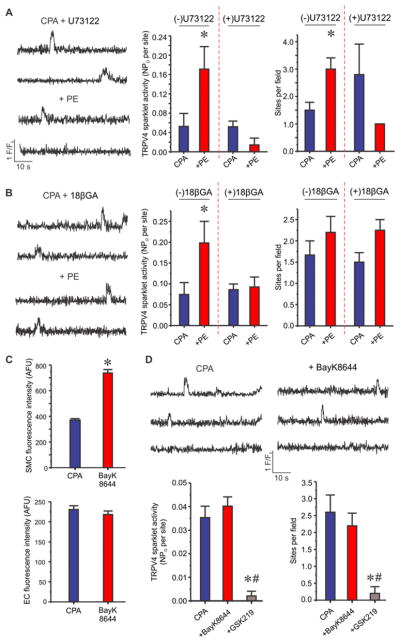
Figure 3. SMC α1AR signaling activates phospholipase C and SMC-EC coupling via MEGJs to stimulate endothelial TRPV4 channels in a Ca2+-independent manner.
Experiments were performed in the presence of CPA (20 μM). (A) Left, representative F/F0 traces for the effect of PE (10 μM) on TRPV4 sparklets in the presence of U73122 (a PLC inhibitor, 1 μM). Right, quantified TRPV4 sparklet activity in response to PE in the absence or presence of U73122 (n=7). * p<0.05 vs. CPA. (B) Left, representative F/F0 traces for the effect of PE (10 μM) on TRPV4 sparklets in the presence of 18βGA (a gap junction uncoupling agent, 30 μM). Right, averaged TRPV4 sparklet activity in response to PE in the absence or presence of 18βGA (n=6). * p<0.05 vs. CPA. (C) Quantified fluorescence intensity indicates whole-cell Ca2+ levels (arbitrary fluorescence units or AFU) in SMCs (top) and ECs (bottom) before and after BayK8644 (a voltage-dependent Ca2+ channel agonist, 1 μM, n=47–50 cells of each). * p<0.05 vs. Baseline. (D) Representative F/F0 traces (top) and quantification (bottom) of averaged TRPV4 sparklet activity in the absence or presence of BayK8644 (1 μM) or BayK8644+GSK219 (100 nM, n=6). TRPV4 Ca2+ sparklet activity is expressed as NPO per site and number of sparklet sites per field (Figure 3A, B, and D). N represents the number of channels at a site and PO is the open state probability of the channels. Data are presented as mean ± SEM.

