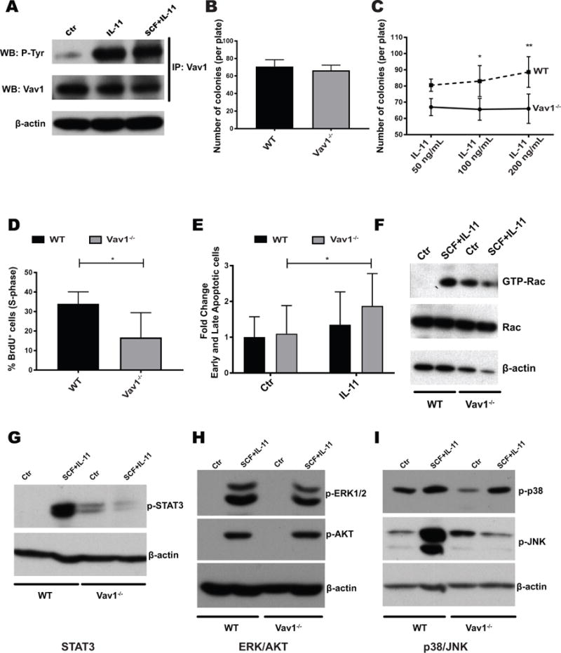
Figure 5. Vav1−/− HSPCs display reduced proliferation, increased apoptosis and abnormal molecular responses to IL-11.

A) Vav1 is phosphorylated upon treatment of HSPCs with IL-11, as demonstrated by immunoprecipitation (IP) with a Vav1-specific antibody, followed by detection with phosphotyrosine antibody (p-Tyr). WT lineage-depleted cells were either left untreated or stimulated with IL-11 alone or SCF and IL-11 (both at 100 ng/mL) for 10 minutes. WB: western blot. B) Number of colonies formed by WT or Vav1−/− BM cells in standard semisolid media (n=12). C) Number of colonies formed by WT or Vav1−/− BM cells in standard semisolid media supplemented with increasing doses of IL-11 (n=4). Error bars represent standard deviation (SD). *P <05; **P <.01. D) Percentage of proliferating cells in liquid cultures established from Vav1−/− and WT HSPCs. Cells were cultured in SCF and IL-11. Bar graphs represent average percentage of cells in S-phase after a 12 h pulse with BrdU (n=3). Error bars represent standard deviation (SD). *P <05. E) Percentage of apoptotic (both early and late) cells per genotype with SCF alone (control) and SCF/IL-11 (IL-11). Bar graphs represent fold change to WT control (n=3). Error bars represent standard deviation (SD). *P <05. F) Levels of active (GTP-bound) and total Rac in WT or Vav1−/− HSPCs. Lineage-depleted cells were starved and left untreated or stimulated with SCF/IL-11. GTP-bound Rac was precipitated with agarose-conjugated PAK1-p21-binding domain (PBD) and detected by western blot. G-I) Activation status of signaling pathways in WT and Vav1−/− lineage-depleted cells by WB with phospho-specific antibodies. Cells were stimulated as in E), and protein lysates were probed with the indicated antibodies. Images display one representative WB of 3 independent experiments.
