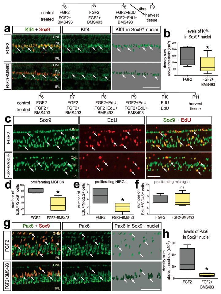
Figure 5.
In the absence of retinal damage, inhibition of RA-signaling with BMS493 suppresses the proliferation of MGPCs in FGF2-treated retinas. a,b: Eyes were injected with FGF2 alone (control) or FGF2+BMS493 (treated) at P6, P7 and P8, and tissues harvested at 4 hrs after the last injection. c–h: Eye were injected with FGF2 alone (control) or FGF2+BMS493 (treated) at P6 and P7, FGF2+EdU or FGF2+BMS439+EdU at P8 and P9, EdU alone at P10, and tissues harvested at P11. Sections of the retina were labeled for Sox9 (red) and Klf4 (green;a); EdU-incorporation (red) and Sox9 (green; c), or Pax6 (green) and Sox9 (red; g). Arrows indicate the nuclei of MGPCs (b, d–f, h). The box plots illustrate the mean, upper extreme, lower extreme, upper quartile and lower quartile (n=7 animals). Significance of difference (*p<0.05) was determined by using a t-test. The calibration bar in panels a, c and g represent 50 μm. Abbreviations: INL – inner nuclear layer, IPL – inner plexiform layer, ONL – outer nuclear layer.

