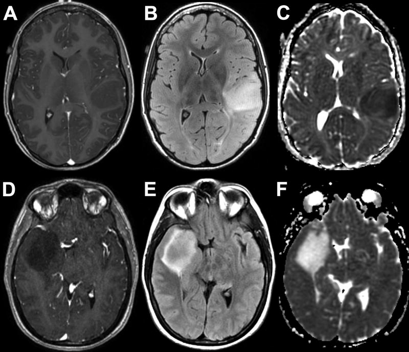
Fig. 1.
Representative MRIs of IDH-wildtype (A–C) and IDH-mutant (D–F) grade II DGs. 23-year-old woman with left frontoparietal opercular non-enhancing (A) and FLAIR hyperintense (B) mass with reduced diffusion on ADC (C). 45-year-old man with righ temporal non-enhancing (D) and FLAIR hyperintense (E) mass with facilitated diffusion on ADC (F).

