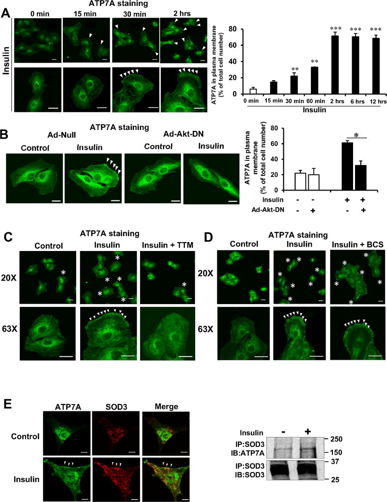
Figure 4. Insulin increases ATP7A translocation to the plasma membrane, where SOD3 colocalizes with ATP7A in an Akt- and Cu-dependent manner in VSMCs.
A–D, RASMs were stimulated with 10 nM of insulin for indicated time (A). RASMs infected with adenovirus expressing Akt-dominant negative (Ad-Akt-DN) or Ad.null (control)(B) or treated with the cell permeable Cu chelator TTM (10 nM, 24 hrs)(C) or treated with cell impermeable Cu chelator, BCS (200 µM, 72 hrs)(D) were stimulated with 10 nM insulin for 2 hrs. These cells were stained with anti-ATP7A antibody, and images were taken at 5 different fields/well from 3 different experiments. Graph represents quantification of plasma membrane ATP7A positive cells. Results are presented as mean± SEM. *p<0.05, **p<0.01, ***p<0.001. E, Immunofluorescence analysis showing the co-localization of ATP7A and SOD3 at plasma membrane (left) or co-immunoprecipitation of SOD3 and ATP7A (right) in human aortic smooth muscle cells (HASMs) stimulated with or without insulin (10 nM, 2 hrs) (n=4). Negative control staining lacking the primary antibodies in each staining is shown in Figure XIV in the online-only Data Supplement. Bar represents 20 µm.

